
Publications
Home
Publications

LUMINA: A Multi-Vendor Mammography Benchmark with Energy Harmonization Protocol
Authors: Hongyi Pan, Gorkem Durak, Halil Ertugrul Aktas, Andrea M Bejar, Baver Tutun, Emre Uysal, Ezgi Bulbul, Mehmet Fatih Dogan, Berrin Erok, Berna Akkus Yildirim, Sukru Mehmet Erturk, Ulas Bagci
CVPR 2026
Publication Year: 2026


Anatomy-Aware Prediction of Bronchoscopic Accessibility from 3D CT
Authors: Linkai Peng, Cuiling Sun, Bin Wang, Jamie Rowell, Catherine Gao, Oyku Ikizgul, Eminenur Sentasci, Andrea Bejar, Halil Ertugrul Aktas, Gorkem Durak, Momen Wahidi, Christopher Kapp, Ulas Bagci
MICCAI 2026
Publication Year:
SRMA-Mamba: Spatial Reverse Mamba Attention Network for Pathological Liver Segmentation in MRI Volumes
Authors: Jun Zeng, Quoc-Huy Trinh, Deepak Ranjan Nayak, Nikhil Tomar Kumar, Ulas Bagci, Debesh Jha
MICCAI 2026
Publication Year: 2026

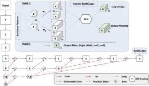
Discrete Cosine Transform Based Decorrelated Attention for Vision Transformers
Authors: Hongyi Pan, Emadeldeen Hamdan, Xin Zhu, Ahmet Enis Cetin, Ulas Bagci
IJCAI-2026
Publication Year: 2026

NeoGaze: Whose Gaze Counts? Expertise, Clinical Information, and Attention in Neonatal Radiology
Authors: Max Bengtsson, Elif Keles, Marouane Tilba, Ulas Bagci
MIPS XXI 2026
Publication Year: 2026

EyeAssist: How Clinical Context Modulates Expert Gaze in Volumetric Reading and Prognostic Decision-Making
Authors: Bin Wang and Ulas Bagci
MIPS XXI 2026 (Medical Image Perception Symposium)
Publication Year: 2026

MSK MRI-Based Personalized Osteochondral Arthroplasty with Tissue-Specific Robotic Resection: A Feasibility Framework
Authors: Sangeun Song , Ulas Bagci , Ramsey Kinney , Jutta Ellermann
iMRI 2026 (interventional MRI symposium)
Publication Year: 2026

PRS-Med: Position Reasoning Segmentation in Medical Imaging
Authors: Quoc-Huy Trinh, Minh-Van Nguyen, Jun Zeng, Debesh Jha, Ulas Bagci
CVPRW 2026 - CV4CLINIC
Publication Year: 2026
CT-DegradBench: A Physics-Informed Benchmark for CT Degradation Detection and Severity Estimation
Authors: Yousra Nabila Taifour, Marouane Tliba, Zuheng Ming, Marie Luong, Nour Aburaed, Aladine Chetouani, Gorkem Durak, Alessandro Bruno, Faouzi Alaya Cheikh, Habib Zaidi, Ulas Bagci, Azeddine Beghdadi
CVPRW (VISION 2026)
Publication Year: 2026
Seeing Through the Tool: A Controlled Benchmark for Occlusion Robustness in Foundation Segmentation Models
Authors: Nhan Ho, Luu Le, Thanh-Huy Nguyen, Thien Nguyen, Xiaofeng Liu, Ulas Bagci
CVPR 2026 - CV4Clinic
Publication Year: 2026
MammoClean: Toward Reproducible and Bias-Aware AI in Mammography through Dataset Harmonization
Authors: Yalda Zafari, Hongyi Pan, Gorkem Durak, Ulas Bagci, Essam A. Rashed, Mohamed Mabrok
IEEE ACCESS
Publication Year: 2026


REN: Anatomically-Informed Mixture-of-Experts for Interstitial Lung Disease Diagnosis
Authors: Alec K. Peltekian, Halil Ertugrul Aktas, Gorkem Durak, Kevin Grudzinski, Bradford C. Bemiss, Carrie Richardson, Jane E. Dematte, G. R. Scott Budinger, Anthony J. Esposito, Alexander Misharin, Alok Choudhary, Ankit Agrawal, Ulas Bagci
IEEE TMI
Publication Year: 2026

Methods of artificial intelligence-assisted infrastructure assessment using mixed reality systems
Authors: Enes Karaaslan, Fikret Necati Catbas, Ulas Bagci
Publication Year: 2026

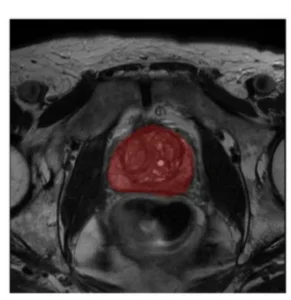
CANCER EXTENT AND CELLULARITY BUT NOT INFLAMMATORY DENSITY ARE ASSOCIATED WITH MRI-INVISIBLE PROSTATE CANCER ON AI-BASED DIGITAL PATHOLOGY DEEP LEARNING MODELS
Authors: Ramin Nateghi, Marina Schnauss, Nicole Handa, Mitchell M Huang, Joseph D Nicolas, Sai Kaushik Shankar Ramesh Kumar, Behtash G Nezami, Ulas Bagci, Edward M Schaeffer, Adam B Murphy, Ximing J Yang, Ashley E Ross, Lee AD Cooper, Hiten D Patel
AUA (American Urology Association) Annual Meeting - 2026
Publication Year: 2026


Triangulating Truth: Experts, Risk Scores, and Machine Learning in Robot-assisted Bronchoscopic Diagnostics
Authors: R Bhargava, C Pfister, Y Liu, JH Rowell, L Peng, J Kim, C Phan, A Demaio, HJ Lee, CR Gilbert, AP Ratwani, JD Duke, T Ferguson, C Argento, SB Smith, M Wayne, J Thiboutot, J Akulian, J Beattie, DH Yu, BJ Seides, MM Wahidi, U Bagci, CA Gao, CM Kapp, BRONCH SMART Study Team
ATS 2026
Publication Year: 2026

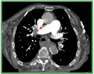
Deep Learning for Pulmonary Embolism Prognosis From CT Imaging: A Multicenter Evaluation
Authors: H Aktas, B Gultekin, V Sharma, A Kirkoglu, O Cetin, K Akin, Z Tuz, N Alibeyoglu, U Durak, K Arisin, R Eren, E Niksarlioglu, N Senkal, M Erelel, A Medetalibeyoglu, S Erturk, G Durak, U Bagci
ATS 2026
Publication Year: 2026

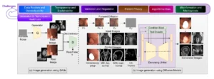
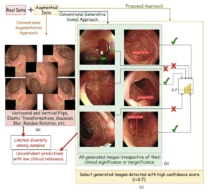
Diverse image generation with diffusion models and cross class label learning for polyp classification
Authors: Vanshali Sharma, Debesh Jha, Manas Kamal Bhuyan, Pradip K Das, Ulas Bagci
Scientific Reports (Nature)
Publication Year: 2026


A Predictive MRI Radiomics Model for Histologic Differentiation in Soft Tissue Sarcomas
Authors: Laetitia Perronne, Nicolò Gennaro, Zuzanna Kobus, Mirinae Seo, Amir A Borhani, Linda Kelahan, Hatice Savas, Ryan Avery, Kamal Subedi, Chase Krumpelman, Gorkem Durak, Ulas Bagci, Akhil Chawla, Borislav Alexiev, Pedro Hermida de Viveiros, Seth Pollack, Yuri S Velichko
Cancers
Publication Year: 2026


Multimodal AI for early prediction of adverse clinical outcomes in acute pancreatitis
Authors: Ahmet Yasin Karkas, Yavuz B Taktak, Burak Gultekin, Ziliang Hong, Halil Ertugrul Aktas, Deniz Seyithanoglu, Timurhan Cebeci, Alper Akin, Ece Elustu, Kerem Arisin, Ali Canturk, Mehmet Ilhan, Naci Senkal, Michael B Wallace, Abraham F Bezuidenhout, Frank H Miller, Alpay Medetalibeyoglu, Mehmet Semih Cakir, Gorkem Durak, Ulas Bagci, Sukru Mehmet Erturk
Abdominal Radiology
Publication Year: 2026


Clinical and Reader Associated Correlates of Clinically Significant Magnetic Resonance Imaging–Invisible Prostate Cancer Based on Negative Magnetic Resonance Imaging
Authors: Marina Schnauss, Nicole Handa, Ramin Nateghi, Ridwan Alam, Jae W Jang, Mitchell M Huang, Aaron Abrams, Clayton Neill, Sai Kaushik Shankar Ramesh Kumar, Nikki Hubbard, Emma McGarrity, Yutai Li, Adam B Murphy, Ulas Bagci, Anugayathri Jawahar, Lee AD Cooper, Ashley E Ross, Hiten D Patel
The Journal of Urology
Publication Year: 2026


Evaluating the Predictive Value of Post-Treatment Superb Microvascular Imaging for Complete Response to Neoadjuvant Chemotherapy in Invasive Breast Cancer
Authors: Rana Gunoz Comert, Ravza Yilmaz, Eda Cingoz, Zuhal Bayramoglu, Aysel Bayram, Baran Mollavelioglu, Mahmut Muslumanoglu, Ulas Bagci
Bioengineering
Publication Year: 2026


Beyond autonomy: why medicine needs artificial intelligence teammates, not artificial intelligence doctors
Authors: G Durak, A Medetalibeyoglu, V Cicek, E Keles, U Bagci
Diagnostic and Interventional Radiology
Publication Year: 2026

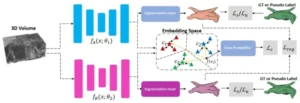
Align then Refine: Text-Guided 3D Prostate Lesion Segmentation
Authors: Cuiling Sun, Linkai Peng, Adam Murphy, Elif Keles, Hiten D Patel, Ashley Ross, Frank Miller, Baris Turkbey, Andrea Mia Bejar, Halil Ertugrul Aktas, Gorkem Durak, Ulas Bagci
IEEE EMBC 2026 (Oral)
Publication Year: 2026
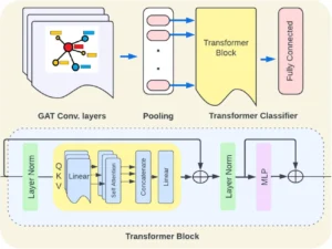
Morphology-Aware KOA Classification: Integrating Graph Priors with Vision Models
Authors: Marouane Tliba, Mohamed Amine Kerkouri, Yassine Nasser, Nour Aburaed, Aladine Chetouani, Ulas Bagci, Rachid Jennane
IEEE EMBC 2026 (Oral)
Publication Year: 2026
Federated Breast Cancer Detection Enhanced by Synthetic Ultrasound Image Augmentation
Authors: Hongyi Pan, Ziliang Hong, Gorkem Durak, Ziyue Xu, Ulas Bagci
IEEE EMBC 2026 (Oral)
Publication Year: 2026
Hybrid Multi-Dimensional MRI Prostate Cancer Detection via Hadamard Network-Based Bias Correction and Residual Networks
Authors: Emadeldeen Hamdan, Gorkem Durak, Muhammed Enes Tasci, Abel Lorente Campos, Aritrick Chatterjee, Roger Engelmann, Gregory Karczma, Aytekin Oto, Ahmet Enis Cetin, Ulas Bagci
IEEE EMBC 2026
Publication Year: 2026

GazeVaLM: A Multi-Observer Eye-Tracking Benchmark for Evaluating Clinical Realism in AI-Generated X-Rays
Authors: David Wong, Zeynep Isik, Bin Wang, Marouane Tliba, Gorkem Durak, Elif Keles, Halil Ertugrul Aktas, Aladine Chetouani, Cagdas Topel, Nicolo Gennaro, Camila Lopes Vendrami, Tugce Agirlar Trabzonlu, Amir Ali Rahsepar, Laetitia Perronne, Matthew Antalek, Onural Ozturk, Gokcan Okur, Andrew C. Gordon, Ayis Pyrros, Frank H. Miller, Amir Borhani, Hatice Savas, Eric Hart, Elizabeth Krupinski, Ulas Bagci
ACM ETRA 2026
Publication Year: 2026
What They Saw, Not Just Where They Looked: Semantic Scanpath Similarity via VLMs and NLP metric
Authors: Mohamed Amine Kerkouri, Marouane Tliba, Bin Wang, Aladine Chetouani, Ulas Bagci, Alessandro Bruno
ACM ETRA 2026
Publication Year: 2026

Ensemble Models for Predicting Treatment Response in Pediatric Low-Grade Glioma Managed with Chemotherapy
Authors: Max Bengtsson, Elif Keles, Angela J. Waanders, Ulas Bagci
IEEE Conference on Artificial Intelligence (CAI) - 2026
Publication Year: 2026

Performance of deep learning‑based segmentation of soft tissuesarcoma by MRI sequence, tumor type and location
Authors: Linkai Peng, Laetitia Perronne, Nicolò Gennaro, Ahmad Pour Rashidi, Zuzanna Kobus, Mirinae Seo, Amir A. Borhani, Linda Kelahan, Kamal Subedi, Hatice Savas, Ryan Avery, Tugce Agirlar Trabzonlu, Chase Krumpelman, Spyridon Bakas, Akhil Chawla, Sean Sachdev, Pedro Hermida de Viveiros, Seth M. Pollack, Ulas Bagci, Yuri S. Velichko
Skeletal Radiology
Publication Year: 2026


Ultra-ECP: Ellipse-Constrained and Point-Robust Foundation Model Adaptation for Fetal Cardiac Ultrasound Segmentation
Authors: Minh HN Le, Khanh TQ Le, Tuan Vinh, Thanh-Huy Nguyen, Han H Huynh, Khoa D Pham, Anh Mai Vu, Hien Quang Kha, Phat Ky Nguyen, Ulas Bagci, Min Xu, Carl Yang, Phat Kim Huynh, Nguyen Quoc Khanh Le
MIDL 2026
Publication Year: 2026
CrossPan: A Comprehensive Benchmark for Cross-Sequence Pancreas MRI Segmentation and Generalization
Authors: Linkai Peng, Cuiling Sun, Zheyuan Zhang, Wanying Dou, Halil Ertugrul Aktas, Andrea M Bejar, Elif Keles, Tamas Gonda, Michael B Wallace, Zongwei Zhou, Gorkem Durak, Rajesh N Keswani, Ulas Bagci
MIDL 2026
Publication Year: 2026
WFM: 3D Wavelet Flow Matching for Ultra-Fast Multi-Modal MRI Synthesis
Authors: Yalcin Tur, Mihajlo Stojkovic, Ulas Bagci
MIDL 2026
Publication Year: 2026

A Multi-center MRI Study with Comprehensive Radiological Analysis of IPMN Risk Stratification with Explainable AI
Authors: H. Aktas, G. Durak, A.M. Bejar, Z. Hong, R. Hendrix, H. Pan, E. Keles, F. Bol, Y. Taktak, C. Topel, Y. Tur, Z. Zhang, Y. Velichko, A. Medetalibeyoglu, S. Erturk, L. Zhao, F.H. Miller, M.B. Wallace, R.N. Keswani, U.Bagci
ESGAR-2026 (Oral)
Publication Year: 2026

IPMN Risk assessment with subregional radiomics and deep learning
Authors: E. Sen Tasi, A.M. Bejar, M. Nelson, H. Aktas, Z. Hong, E. Keles, M. Tasci, S. Kayali, F. Farahmand, F. Miller, M.B. Wallace, R. Keswani, G. Durak, U. Bagci
ESGAR-2026
Publication Year: 2026

MRI-Based Radiomics Model for Risk Stratification of Main Duct IPMNs: Toward Reducing Unnecessary Pancreatic Surgery
Authors: Ertugrul Aktas et al.
DDW 2026
Publication Year: 2026
Acute Pancreatitis Outcome Prediction with Deep Learning and Radiomics
Authors: Ertugrul Aktas et al
DDW 2026
Publication Year: 2026
Noninvasive Imaging Biomarkers of Cirrhosis: Functional Liver Imaging Score and Liver Surface Modularity Correlations with MELD-NA
Authors: Enes Tasci, et al.
DDW 2026
Publication Year: 2026

Multi-Center Radiomics-Deep lEarning Fusion Model for Stratifying IPMN Malignancy Risk on MRI
Authors: Bejar, A., et al
ISMRM 2026
Publication Year: 2026
Liver Cirrhosis Visual Severity Estimation From MRI With Deep Learning
Authors: Aktas, H.E., et al
ISMRM 2026
Publication Year: 2026
VHU-Net: Variational Hadamard U-Net for Body MRI Bias Field Correction
Authors: Zhu, X., et al
ISMRM 2026
Publication Year: 2026
Imaging Biomarkers of Cirrhosis: Functional Liver Imaging Score and Liver Surface Nodularity Correlations with MELD-Na
Authors: Tasci, M.E, et al.
ISMRM 2026
Publication Year: 2026
In-depth Radiological and Explainability Analysis of Deep Learning Predictions for IPMN Risk Stratification on MRI
Authors: Aktas, E. et al
ISMRM 2026
Publication Year: 2026

The 2024 Brain Tumor Segmentation Challenge Meningioma Radiotherapy (BraTS-MEN-RT) dataset
Authors: Dominic LaBella, Katherine Schumacher, Michael Mix, Kevin Leu, Shan McBurney-Lin, Pierre Nedelec, Javier Villanueva-Meyer, David R Raleigh, Jonathan Shapey, Tom Vercauteren, Kazumi Chia, Marina Ivory, Theodore Barfoot, Omar Al-Salihi, Justin Leu, Lia M Halasz, Yury Velichko, Chunhao Wang, John P Kirkpatrick, Scott R Floyd, Zachary J Reitman, Trey C Mullikin, Eugene J Vaios, Ulas Bagci, Sean Sachdev, Jona A Hattangadi-Gluth, Tyler M Seibert, Nikdokht Farid, Connor Puett, Matthew W Pease, Kevin Shiue, Syed M Anwar, Shahriar Faghani, Peter Taylor, Pranav Warman, Jake Albrecht, András Jakab, Mana Moassefi, Verena Chung, Rong Chai, Alejandro Aristizabal, Alexandros Karargyris, Hasan Kassem, Sarthak Pati, Micah Sheller, Nazanin Maleki, Rachit Saluja, Florian Kofler, Christopher G Schwarz, Philipp Lohmann, Phillipp Vollmuth, Louis Gagnon, Maruf Adewole, Li Hongwei B, Anahita Fathi Kazerooni, Nourel H Tahon, Udunna Anazodo, Ahmed W Moawad, Bjoern Menze, Marius G Linguraru, Mariam Aboian, Benedikt Wiestler, Ujjwal Baid, Gian-Marco Conte, Andreas M Rauschecker, Ayman Nada, Aly H Abayazeed, Raymond Huang, Maria Correia de Verdier, Jeffrey D Rudie, Spyridon Bakas, Evan Calabrese
Nature Scientific Data
Publication Year: 2026


AI-Driven Multimodal MRI Framework for Automated Staging of Laryngeal Cancer
Authors: F Bol, M Iren, M Mureva, T-H Nguyen, T Nguyen, E Aktas, B Yildirim, U Bagci, B Atasoy
ECR 2026
Publication Year: 2026


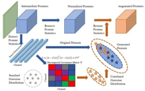
Domain-invariant Mixed-domain Semi-supervised Medical Image Segmentation with Clustered Maximum Mean Discrepancy Alignment
Authors: Ba-Thinh Lam, Thanh-Huy Nguyen, Hoang-Thien Nguyen, Quang-Khai Bui-Tran, Nguyen Lan Vi Vu, Phat K. Huynh, Ulas Bagci, Min Xu
IEEE ICASSP 2026
Publication Year: 2026
Aligning What You Separate
Authors: Quang-Khai Bui-Tran, Thanh-Huy Nguyen, Hoang-Thien Nguyen, Ba-Thinh Lam, Nguyen Lan Vi Vu, Phat K. Huynh, Ulas Bagci, Min Xu
IEEE ICASSP 2026
Publication Year: 2026

VHU-Net: Variational Hadamard U-Net for Body MRI Bias Field Correction
Authors: Xin Zhu, Ahmet Enis Cetin , Gorkem Durak, Batuhan Gundogdu, Ziliang Hong, Hongyi Pan, Ertugrul Aktas, Elif Keles, Hatice Savas, Aytekin Oto, Hiten Patel, Adam B. Murphy, Ashley Ross, Frank Miller, Baris Turkbey, Ulas Bagci
Medical Image Analysis
Publication Year: 2026
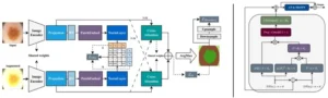
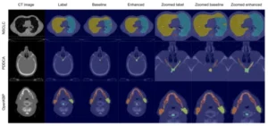
Coronary artery segmentation in non-contrast cardiac CT using anatomy-informed contrastive learning and synthetic data
Authors: Jinkui Hao, Xiaoyi He, Gorkem Durak, Halil Ertugrul Aktas, Ulas Bagci, Nilay S Shah, Bo Zhou
Physics in Medicine and Biology
Publication Year: 2026


Robust White Blood Cell Classification with Stain-Normalized Decoupled Learning and Ensembling
Authors: L Le, HL Cao, HH Pham, TH Nguyen, U Bagci
IEEE ISBI 2026
Publication Year: 2026

Handling supervision scarcity in chest x-ray classification: Long-tailed and zero-shot learning
Authors: Ha-Hieu Pham, Hai-Dang Nguyen, Thanh-Huy Nguyen, Min Xu, Ulas Bagci, Trung-Nghia Le, Huy-Hieu Pham
IEEE ISBI 2026
Publication Year: 2026
Scribble-Supervised Medical Image Segmentation with Dynamic Teacher Switching and Hierarchical Consistency
Authors: Huy Thanh Nguyen; Loc Hoang Cao and Dat Tien Chung; Anh Mai Vu; Thanh-Minh Nguyen; Minh Huu Nhat Le; Phat Kim Huynh ; Ulas Bagci
IEEE ISBI 2026
Publication Year: 2026
Upstream Probabilistic Meta-Imputation for Multimodal Pediatric Pancreatitis Classification
Authors: Max A. Nelson, Elif Keles, Eminenur Sen Tasci, Merve Yazol, Halil Ertugrul Aktas, Ziliang Hong, Andrea Mia Bejar, Gorkem Durak, Oznur Leman Boyunaga, Ulas Bagci
IEEE ISBI 2026
Publication Year: 2026
CTest-Metric: A Unified Framework to Assess Clinical Validity of Metrics for CT Report Generation
Authors: Vanshali Sharma, Andrea Mia Bejar, Gorkem Durak, Ulas Bagci
IEEE ISBI 2026
Publication Year: 2026
Multi-center evaluation of radiomics and deep learning to stratify malignancy risk of IPMNs
Authors: Andrea M Bejar, Maria Jaramillo Gonzalez, Ziliang Hong, Gorkem Durak, Elif Keles, Halil Ertugrul Aktas, Zheyuan Zhang, Hongyi Pan, Zeynep Sue Jozwiak, Fergan Bol, Lili Zhao, Chao Chen, Concetto Spampinato, Alpay Medetalibeyoglu, Sukru Mehmet Erturk, Gulbiz Dagoglu Kartal, Yury Velichko, Emil Agarunov, Ziyue Xu, Sachin Jambawalikar, Ivo G Schoots, Marco J Bruno, Chenchang Huang, Tamas Gonda, Candice Bolan, Frank H Miller, Michael B Wallace, Rajesh N Keswani, Pallavi Tiwari & Ulas Bagci
Abdominal Radiology
Publication Year: 2026

Comparing Dermatologist and Artificial Intelligence Heat Maps in Dermoscopic Image Analysis via Eye Tracking
Authors:
Journal of the American Academy of Dermatology - 2026
Publication Year: 2026


Large language models standardize the interpretation of complex oncology guidelines for brain metastases
Authors: Berna Akkus Yildirim, Baver Tutun, Gorkem Durak, Emre Batuhan Yildirim, Emre Uysal, Sukru Mehmet Erturk, Ulas Bagci
Communications Medicine (Nature)
Publication Year: 2026


AI in the Hot Seat: Head-to-Head Comparison of Large Language Models and Cardiologists in Emergency Scenarios
Authors: Vedat Cicek, Lili Zhao, Yalcin Tur, Ahmet Oz, Sahhan Kilic, Gorkem Durak, Faysal Saylik, Mert Ilker Hayiroglu, Tufan Cinar, Ulas Bagci
Medical Sciences
Publication Year: 2026
UP2D: Uncertainty-aware progressive pseudo-label denoising for source-free domain adaptive medical image segmentation
Authors: Thanh-Huy Nguyen, Quang-Khai Bui-Tran, Manh D Ho, Thinh B Lam, Vi Vu, Hoang-Thien Nguyen, Phat Huynh, Ulas Bagci
Neurocomputing
Publication Year: 2026

Prototype Learning for Out-of-Distribution Polyp Segmentation
Authors: Nikhil Kumar Tomar, Debesh Jha, Ulas Bagci
AAAI 2026 (AIMedHealth26 Workshop)
Publication Year: 2026

IPMN Risk Assessment with Subregional Radiomics and Deep Learning
Authors: Andrea Mia Bejar, Eminenur Sen Tasci, Max Nelson, Halil Ertugrul Aktas, Katie Wu, Ziliang Hong, Elif Keles, Muhammed Enes Tasci, Frank H Miller, Michael B Wallace, Rajesh N Keswani, Gorkem Durak, Ulas Bagci
MAYO CLINIC INNOVATIONS IN GASTROENTEROLOGY AND HEPATOLOGY 2026: ARTIFICIAL INTELLIGENCE AND BEYOND
Publication Year: 2026
In-depth Radiological and Explainability Analysis of Deep Learning
Authors: Halil Ertugrul Aktas, Andrea Mia Bejar, Ziliang Hong, Rutger Hendrix, Hongyi Pan, Elif Keles, Fergan Bol, Yavuz Bahadir Taktak, Yuri Velichko, Alpay Medetalibeyoglu, Sukru Mehmet Erturk, Lili Zhao, Frank H. Miller, Michael Wallace, Rajesh N. Keswani, Gorkem Durak, and Ulas Bagci
Mayo Clinic Innovations in Gastroenterology and Hepatology 2026: Artificial Intelligence and Beyond
Publication Year: 2026
Functional MRI Scoring as a Noninvasive Method to Predict Hepatic Functional Reserve
Authors: G. Durak, Y.B. Taktak, B. Gültekin, S. Bayrak, E. Özkan, Ö. Ikizgül, E. Aktas, M. Ertürk, A. Medetalibeyoglu, U. Bagci
ASC 2026
Publication Year: 2026


Predicting Pulmonary Fibrosis Risk in Post-COVID Patients using Deep Learning and Radiomics on CT
Authors: Wanying Dou, Andrea Bejar, Halil Ertugrul Aktas, Gorkem Durak, Elif Keles, Drew Torigian, Jayaram Udupa, Ulas Bagci
ARRS 2026
Publication Year: 2026
Validating Synthetically Generated Medical Scans through Human Perceptual Assessment & Eye-Tracking
Authors: David Wong, Bin Wang, Gorkem Durak, Halil Ertugrul Aktas, Marouane Tliba, Hatice Savas, Elizabeth Krupinski, Ulas Bagci
ARRS 2026
Publication Year: 2026
Detecting Metaplastic Histology in Triple-Negative Breast Cancer with Machine Learning
Authors: Rana Gunoz Comert, Gorkem Durak, Ravza Yilmaz, Halil Ertugrul Aktas, Zeynep Tuz, Jun Zeng , Sukru Mehmet Erturk, Ulas Bagci
ARRS 2026
Publication Year: 2026
Improved Segmentation of Polyps and Visual Explainability Analysis
Authors: Akwasi Asare, Thanh-Huy Nguyen, Ulas Bagci
IEEE ACDSA 2026
Publication Year: 2026


ROLE OF IMAGING IN PANCREATIC CANCER SCREENING FOR HIGH-RISK POPULATION WITH GENETIC AND HEREDITARY CAUSES LINKED TO SPECIFIC GENETIC SYNDROMES OR MUTATIONS
Authors: Pardeep K. Mittal, MD, Srinivasa R. Prasad, MD, Anil K. Dasyam, MD, Venkata S. Katabathina, MD, Courtney C. Moreno, MD, Camila Lopes Vendrami, MD, Ulas Bagci, MSc, PhD, Frank H. Miller
RSNA 2025
Publication Year: 2025
Outperforming State-of-the-Art in Pediatric Brain Tumor Segmentation with a Dual NNU-Net Model Based on Radiological Reasoning
Authors: Elif Keles, Max Bengtsson, Gorkem Durak, Syed M. Anwar, Yuri Velichko, Marius G. Linguraru, Angela Waanders, Ulas Bagci
RSNA 2025
Publication Year: 2025
A PREDICTIVE MRI RADIOMICS MODEL FOR IMPROVED SOFT TISSUE SARCOMA SUBTYPE DIFFERENTIATION
Authors: Laetitia Perronne, MD, MSc, Nicolo Gennaro, MD, Mirinae Seo, MD, Zuzanna Kobus, MD, Linkai Peng, BEng, Gorkem Durak, MD, Ulas Bagci, MSc, PhD, Amir Borhani, MD, Hatice Savas, MD, Linda C. Kelahan, MD, Kamal Subedi, MD, Ryan J. Avery, MD, Tugce Agirlar Trabzonlu, MD, Chase S. Krumpelman, MD, PhD, Seth Pollack, MD, Pedro Hermida De Viveiros, Yuri Velichko, PhD
RSNA 2025
Publication Year: 2025
Foundational Tumor Segmentation Models
Authors: Sirui Li, Linkai Peng, Halil Ertugrul Aktas, Zheyuan Zhang, Gorkem Durak, Ulas Bagci,
RSNA 2025
Publication Year: 2025
Multi-Center Radiomics–Deep Learning Fusion Model for Stratifying IPMN Malignancy Risk on MRI
Authors: Andrea Bejar, Ziliang Hong, Gorkem Durak, Elif Keles, Halil Ertugrul Aktas, Zheyuan Zhang, Hongyi Pan, Fergan Bol, Chao Chen, Concetto Spampinato, Alpay Medetalibeyoglu, Sukru Mehmet Erturk, Gulbiz M. Kartal, Yuri Velichko, Emil Agarunov, Ziyue Xu, Sachin Jambawalikar, Ivo Schoots, Marco Bruno, Tamas Gonda, Candice W. Bolan, Frank H. Miller, Rajesh Keswani, Pallavi Tiwari, Ulas Bagci
RSNA 2025
Publication Year: 2025
Hybrid Intelligence in Oncology: Comparing Human Experts and Large Language Models in Interpreting Complex Cancer Treatment Guidelines
Authors: Berna Yildirim, Baver Bunyamin Tutun, Gorkem Durak, Emre Batuhan Yildirim, Emre Uysal, Sukru Mehmet Erturk, Ulas Bagci,
RSNA 2025
Publication Year: 2025
IPMN Malignancy Risk Assessment Using MRI Under Federated Learning Paradigm
Authors: Halil Ertugrul Aktas, Andrea Bejar, Ziliang Hong, Hongyi Pan, Gorkem Durak, Elif Keles, Alpay Medetalibeyoglu, Zheyuan Zhang, Yuri Velichko, Concetto Spampinato, Ivo Schoots, Marco Bruno, Pallavi Tiwari, Candice W. Bolan, Tamas Gonda, Frank H. Miller, Rajesh Keswani, Ziyue Xu, Ulas Bagci
RSNA 2025
Publication Year: 2025
Imaging Biomarkers for Pancreatic Adenocarcinoma Risk Stratification in Incidental Pancreatic Cysts: A Multi-Hospital Cohort Study
Authors: Andrea Bejar, Gorkem Durak, Frank H. Miller, Ulas Bagci, Rajesh Keswani
RSNA 2025
Publication Year: 2025
Automated 3D CT Report Generation Using LLM with Expert-Informed Assessment
Authors: Vanshali Sharma, Andrea Bejar, Wanying Dou, Gorkem Durak, Ulas Bagci
RSNA 2025
Publication Year: 2025
Pediatric Pancreas Segmentation from MRI Scans with Deep Learning
Authors: Elif Keles, Merve Yazol, Gorkem Durak, Ziliang Hong, Halil Ertugrul Aktas, Zheyuan Zhang, Linkai Peng, Cemal Yazici, Ulas Bagci
RSNA 2025
Publication Year: 2025

Generative AI and Foundation Models in Radiology: Applications, Opportunities, and Potential Challenges
Authors: Neda Tavakoli, Zahra Shakeri, Vrushab Gowda, Konrad Samsel, Arash Bedayat, Ahmadreza Ghasemiesfe, Ulas Bagci, Albert Hsiao, Tim Leiner, James Carr, Daniel Kim, Amir Ali Rahsepar
RADIOLOGY
Publication Year: 2025

SAM-guided prompt learning for Multiple Sclerosis lesion segmentation
Authors: Federica Proietto Salanitri, Giovanni Bellitto, Salvatore Calcagno, Ulas Bagci, Concetto Spampinato, Manuela Pennisi
Pattern Recognition Letters
Publication Year: 2025
DuetMatch: Harmonizing semi-supervised brain MRI segmentation via decoupled branch optimization
Authors: Thanh-Huy Nguyen, Hoang-Thien Nguyen, Vi Vu, Ba-Thinh Lam, Phat Huynh, Tianyang Wang, Xingjian Li, Ulas Bagci, Min Xu
Computerized Medical Imaging and Graphics
Publication Year: 2025
AdverIN: Monotonic Adversarial Intensity Attack for Domain Generalization in Medical Image Segmentation
Authors: Zheyuan Zhang, Bin Wang, Lanhong Yao, Elif Keles, Debesh Jha, Matthew Antalek, Gorkem Durak, Alpay Medetalibeyoglu, Concetto Spampinato, Baris Turkbey, Boqing Gong, Ulas Bagci
Medical Image Analysis
Publication Year: 2025

Label-Efficient Cross-Modality Generalization for Liver Segmentation in Multi-Phase MRI
Authors: Quang-Khai Bui-Tran, Minh-Toan Dinh, Thanh-Huy Nguyen, Ba-Thinh Lam, Mai-Anh Vu, Ulas Bagci
CARE 2025 Challenge - MICCAI
Publication Year: 2025
Pancreas Part Segmentation under Federated Learning Paradigm
Authors: Ziliang Hong, Halil Ertugrul Aktas, Andrea Mia Bejar, Katherine Wu, Hongyi Pan, Gorkem Durak, Zheyuan Zhang, Sait Kayali, Temel Tirkes, Federica Proietto Salanitri, Concetto Spampinato, Michael Goggins, Tamas Gonda, Candice Bolan, Raj Keswani, Frank Miller, Michael Wallace, Ulas Bagci
Predictive Intelligence in Medicine (PRIME)-MICCAI
Publication Year: 2025
Learning Disentangled Stain and Structural Representations for Semi-Supervised Histopathology Segmentation
Authors: Ha-Hieu Pham, Nguyen Lan Vi Vu, Thanh-Huy Nguyen, Ulas Bagci, Min Xu, Trung-Nghia Le, Huy-Hieu Pham
MICCAI 2025 (COMPAYL Workshop)
Publication Year: 2025
Imaging-Based Mortality Prediction in Patients with Systemic Sclerosis
Authors: Alec K Peltekian, Karolina Senkow, Gorkem Durak, Kevin M Grudzinski, Bradford C Bemiss, Jane E Dematte, Carrie Richardson, Nikolay S Markov, Mary Carns, Kathleen Aren, Alexandra Soriano, Matthew Dapas, Harris Perlman, Aaron Gundersheimer, Kavitha C Selvan, John Varga, Monique Hinchcliff, Krishnan Warrior, Catherine A Gao, Richard G Wunderink, GR Budinger, Alok N Choudhary, Anthony J Esposito, Alexander V Misharin, Ankit Agrawal, Ulas Bagci
Predictive Intelligence in Medicine (PRIME) - MICCAI
Publication Year: 2025
GMAT: Grounded Multi-agent Clinical Description Generation for Text Encoder in Vision-Language MIL for Whole Slide Image Classification
Authors: Ngoc Bui Lam Quang, Nam Le Nguyen Binh, Thanh-Huy Nguyen, Le Thien Phuc Nguyen, Quan Nguyen, Ulas Bagci
International Workshop on Emerging LLM/LMM Applications in Medical Imaging - MICCAI
Publication Year: 2025
Radiomics for Detecting Metaplastic Histology in Triple-Negative Breast Cancer: A Step Towards Personalized Therapy
Authors: Rana Gunoz Comert, Gorkem Durak, Ravza Yilmaz, Halil Ertugrul Aktas, Zeynep Tuz, Hongyi Pan, Jun Zeng, Aysel Bayram, Baran Mollavelioglu, Sukru Mehmet Erturk, Ulas Bagci
Bioengineering
Publication Year: 2025

ViCTr: Vital Consistency Transfer for Pathology Aware Image Synthesis
Authors: Onkar Susladkar, Gayatri Deshmukh, Yalcin Tur, Gorkem Durak, Ulas Bagci
IEEE ICCV 2025
Publication Year: 2025
VideoAds for Fast-Paced Video Understanding: Where Opensource Foundation Models Beat GPT-4o & Gemini-1.5 Pro
Authors: Zheyuan Zhang, Monica Dou, Linkai Peng, Hongyi Pan, Ulas Bagci, Boqing Gong
IEEE ICCV 2025
Publication Year: 2025
Describe Anything Model for Visual Question Answering on Text-rich Images
Authors: Yen-Linh Vu, Dinh-Thang Duong, Truong-Binh Duong, Anh-Khoi Nguyen, Thanh-Huy Nguyen, Le Thien Phuc Nguyen, Jianhua Xing, Xingjian Li, Tianyang Wang, Ulas Bagci, Min Xu
ICCV 2025 - VisionDocs
Publication Year: 2025
TAGS: 3D Tumor-Adaptive Guidance for SAM
Authors: Sirui Li, Linkai Peng, Zheyuan Zhang, Gorkem Durak, Ulas Bagci
ICCVW 2025 (APAH workshop)
Publication Year: 2025

Order-aware Interactive Segmentation
Authors: Bin Wang, Anwesa Choudhuri, Meng Zheng, Zhongpai Gao, Benjamin Planche, Andong Deng, Qin Liu, Terrence Chen, Ulas Bagci, Ziyan Wu
ICLR 2025
Publication Year: 2025


Eye Tracking in Cardiology
Authors: Vedat Cicek, Ulas Bagci
Current Opinion in Biomedical Engineering
Publication Year: 2025

Unsupervised Clustering Successfully Predicts Prognosis in NSCLC Brain Metastasis Cohorts
Authors: Emre Uysal, Gorkem Durak, Ayse Kotek Sedef, Ulas Bagci, Tanju Berber, Necla Gurdal, Berna Akkus Yildirim
Diagnostics
Publication Year: 2025
Large Scale MRI Collection and Segmentation of Cirrhotic Liver
Authors: Debesh Jha, Onkar Kishor Susladkar, Vandan Gorade, Elif Keles, Matthew Antalek, Deniz Seyithanoglu, Timurhan Cebeci, Halil Ertugrul Aktas, Gulbiz Dagoglu Kartal, Sabahattin Kaymakoglu, Sukru Mehmet Erturk, Yuri Velichko, Daniela P. Ladner, Amir A. Borhani, Alpay Medetalibeyoglu, Gorkem Durak & Ulas Bagci
Nature Scientific Data - 2025
Publication Year: 2025

Generative AI and unstructured audio data for precision public health
Authors: James Anibal, Adam Landa, Hang Nguyen, Veronica Daoud, Tram Le, Hannah Huth, Miranda Song, Alec Peltekian, Ashley Shin, Lindsey Hazen, Anna Christou, Jocelyne Rivera, Robert Morhard, Jacqueline Brenner, Ulas Bagci, Ming Li, Yael Bensoussan, David Clifton & Bradford Wood
Nature (npj) Health Systems - 2025
Publication Year: 2025


Eyes Tell the Truth: GazeVal Highlights Shortcomings of Generative AI in Medical Imaging
Authors: David Wong*, Bin Wang*, Gorkem Durak, Marouane Tliba, Akshay Chaudhari, Aladine Chetouani, Ahmet Enis Cetin, Cagdas Topel, Nicolo Gennaro, Camila Lopes Vendrami, Tugce Agirlar Trabzonlu, Amir Ali Rahsepar, Laetitia Perronne, Matthew Antalek, Onural Ozturk, Gokcan Okur, Andrew C Gordon, Ayis Pyrros, Frank H Miller, Amir Borhani, Hatice Savas, Eric Hart, Drew Torigian, Jayaram K Udupa, Elizabeth Krupinski, Ulas Bagci
CVPRW 2025
Publication Year: 2025


MedDelinea: Scalable and Efficient Medical Image Segmentation via Controllable Diffusion Transformers
Authors: Gayatri Deshmukh, Onkar Kishor Susladkar, Debesh Jha, Elif Keles, Halil Ertugrul Aktas, Daniela P Ladner, Amir A Borhani, Gorkem Durak, Ulas Bagci
MIDL 2025
Publication Year: 2025

Machine learning based clinical decision tool to predict acute kidney injury and survival in therapeutic hypothermia treated neonates
Authors: Elif Keles, Syed Yaseen Ali, Pia Wintermark, Pieter Annaert, Floris Groenendaal, Suzan Şahin, Mehmet Yekta Öncel, Didem Armangil, Esin Koc, Malcolm R. Battin, Alistair J. Gunn, Adam Frymoyer, Valerie Chock, Djalila Mekahli, John van den Anker, Anne Smits, Karel Allegaert & Ulas Bagci
Nature Scientific Reports - 2025
Publication Year: 2025

Negative Predictive Value of a Prostate MRI in Black Men: Implications for Biopsy Decision-Making
Authors: Sarah Sandlow, Samuel Carbunaru, Zequn Sun, Bernice Ofori, Yingzhe Liu, Eric Li, Courtney MP Hollowell, Patricia Vidal, Kent T Perry Jr, Hiten D Patel, Ryan W Dobbs, David D Casalino, Cecil G Wood III, Anugayathri Jawahar, Channa Amarasekera, Jeanne M Horowitz, Frank H Miller, Emrah Gumusgoz, Ximing J Yang, Bonnie Choy, Frances Manosca, Ulas Bagci, Edward M Schaeffer, Peter Gann, Dustin D French, Ashley E Ross, Shilajit D Kundu, Adam B Murphy
Journal of Urology - 2025
Publication Year: 2025
Anatomy-derived 3D Aortic Hemodynamics Using Fluid Physics–informed Deep Learning
Authors: Haben Berhane, Anthony Maroun, David Dushfunian, Justin Baraboo, Gabriela Martinez, Tyler Jacobson, Ulas Bagci, Bradley D. Allen, Michael Markl
Radiology - 2025
Publication Year: 2025
Validating polyp and instrument segmentation methods in colonoscopy through Medico 2020 and MedAI 2021 Challenges
Authors: Debesh Jha, Vanshali Sharma, Debapriya Banik, Debayan Bhattacharya, Kaushiki Roy, Steven A. Hicks, Nikhil Kumar Tomar, Vajira Thambawita, Adrian Krenzer, Ge-Peng Ji, Sahadev Poudel, George Batchkala, Saruar Alam, Awadelrahman M. A. Ahmed, Quoc-Huy Trinh, Zeshan Khan, Tien-Phat Nguyen, Shruti Shrestha, Sabari Nathan, Jeonghwan Gwak, Ritika K. Jha, Zheyuan Zhang, Alexander Schlaefer, Debotosh Bhattacharjee, M.K. Bhuyan, Pradip K. Das, Deng-Ping Fan, Sravanthi Parsa, Sharib Ali, Michael A. Riegler, Pål Halvorsen, Thomas De Lange, Ulas Bagci
Medical Image Analysis - 2025
Publication Year: 2025

Physics-Informed Autoencoder for Prostate Tissue Microstructure Profiling with Hybrid Multidimensional MRI
Authors: Batuhan Gundogdu, Aritrick Chatterjee, Milica Medved, Ulas Bagci, Gregory S Karczmar, Aytekin Oto
Radiology AI - 2025
Publication Year: 2025

Advances in MRI in the Evaluation of Chronic Pancreatitis and the less Common Forms of Pancreatitis
Authors: Camila Lopes Vendrami, Nancy Hammond, Pardeep Mittal, Rajesh N. Keswani, Ulas Bagci, Frank H. Miller
Advances in Clinical Radiology - 2025
Publication Year: 2025

Radiologic and Clinical Correlates of Long-Term Post-COVID-19 Pulmonary Sequelae
Authors: Gorkem Durak, Kaan Akin, Okan Cetin, Emre Uysal, Halil Ertugrul Aktas, Ulku Durak, Ahmet Yasin Karkas, Naci Senkal, Hatice Savas, Atadan Tunaci, Alpay Medetalibeyoglu, Ulas Bagci and Sukru Mehmet Erturk
Journal of Clinical Medicine - 2025
Publication Year: 2025

Pediatric Pancreas Segmentation from MRI Scans with Deep Learning
Authors: Elif Keles, Merve Yazol, Gorkem Durak, Ziliang Hong, Halil Ertugrul Aktas, Zheyuan Zhang, Linkai Peng, Onkar Susladkar, Necati Guzelyel, Oznur Leman Boyunaga, Cemal Yazici, Mark Lowe, Aliye Uc, Ulas Bagci
Pancreatology - 2025
Publication Year: 2025


Position of artificial intelligence in healthcare and future perspective
Authors: Vedat Cicek and Ulas Bagci
Artificial Intelligence in Medicine (Elsevier) - 2025
Publication Year: 2025


Gaze-Guided Medical Image Segmentation: A Training-Free Approach Using SAM Foundation Model
Authors: Giulia Gabriëlla Pötgens, Rutger Hendrix, Cian Scannell, Ulas Bagci, Concetto Spampinato, Federica Proietto Salanitri
IEEE EMBC 2025
Publication Year: 2025


Shifts in Doctors’ Eye Movements Between Real and AI-Generated Medical Images
Authors: David Wong, Bin Wang, et al.
ETRA 2025
Publication Year: 2025


Impact of AI-Generated ADC Maps on Computer-Aided Diagnosis of Prostate Cancer: A Feasibility Study
Authors: Kutsev B. Ozyoruk, Stephanie A. Harmon, Enis C. Yilmaz, David G. Gelikman, Ulas Bagci, Benjamin D. Simon, Maria J. Merino, Rosina Lis, Sandeep Gurram, Bradford J. Wood, Peter A. Pinto, Peter L. Choyke , Baris Turkbey
Academic Radiology (Elsevier) - 2025
Publication Year: 2025

Current applications of radiomics in the assessment of tumor microenvironment of hepatocellular carcinoma
Authors: Junghwa Choi et al
Abdominal Radiology - 2025
Publication Year: 2025


Uncertainty-Guided Cross Attention Ensemble Mean Teacher for Semi-supervised Medical Image Segmentation
Authors: Meghana Karri, Amit Soni Arya, Koushik Biswas, Nicolo Gennaro, Vedat Cicek, Gorkem Durak, Yury Velichko, Ulas Bagci
IEEE/CVF WACV 2025
Publication Year: 2025


Frequency-Domain Refinement of Vision Transformers for Robust Medical Image Segmentation under Degradation
Authors: Sanaz Karimijafarbigloo, Sina Ghorbani Kolahi, Reza Azad, Ulas Bagci, Dorit Merhof
IEEE/CVF WACV 2025
Publication Year: 2025


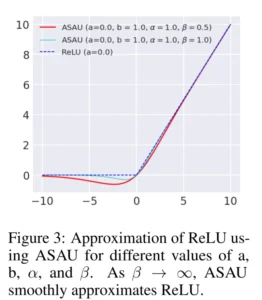
Optimizing Neural Network Effectiveness via Non-monotonicity Refinement
Authors: Koushik Biswas, Amit Reza, Meghana Karri, Debesh Jha, Hongyi Pan, Nikhil Tomar, Aliza Subedi, Smriti Regmi, Ulas Bagci
IEEE/CVF WACV 2025
Publication Year: 2025

Rosette Trajectory MRI Reconstruction with Vision Transformers
Authors: M. Fikret Yalcinbas, Cengizhan Ozturk, Onur Ozyurt, Uzay E. Emir and Ulas Bagci
Tomography - 2025
Publication Year: 2025


A Review of Ultrasonic Testing and Evaluation Methods with Applications in Civil NDT/E
Authors: Inad Alqurashi, Ninel Alver, Ulas Bagci and Fikret Necati Catbas
Journal of Nondestructive Evaluation
Publication Year: 2025

Enhancing Shared Decision-Making in Cardiology with Artificial Intelligence
Authors: Vedat Cicek and Ulas Bagci
Balkan Med J - 2025
Publication Year: 2025


Radiomics-Based Machine Learning Models Improve Acute Pancreatitis Severity Prediction
Authors: Ahmet Yasin Karkas et al.
AI - 2025
Publication Year: 2025


Ethical Framework for Responsible Foundational Models in Medical Imaging
Authors: Debesh Jha , Gorkem Durak , Abhijit Das , Jasmer Sanjotra , Onkar Susladkar , Suramyaa Sarkar , Ashish Rauniyar , Nikhil Kumar Tomar , Linkai Peng , Sirui Li , Koushik Biswas , Ertugrul Aktas , Elif Keles , Matthew Antalek , Zheyuan Zhang , Bin Wang , Xin Zhu , Hongyi Pan , Deniz Seyithanoglu , Alpay Medetalibeyoglu , Vanshali Sharma , Vedat Çiçek , Amir Ali Rahsapar , Rutger Hendrix , A. Enis Cetin , Bulent Aydogan , Mohamed Abazeed , Frank H Miller , Rajesh N Keswani , Hatice Savas , Sachin Jambawalikar , Daniela P Ladner , Amir Ali Borhani , Concetto Spampinato , Michael B Wallace and Ulas Bagci
Frontiers in Medicine
Publication Year: 2025

Paradoxical Response to Neoadjuvant Therapy in Undifferentiated Pleomorphic Sarcoma: Increased TumorSize on MRI Associated with Favorable Pathology
Authors: Mariam H. Goreish
Cancers
Publication Year: 2025

Predicting Risk of Pulmonary Fibrosis Formation in PASC Patients
Authors: Wanying Dou, Gorkem Durak, Koushik Biswas, Ziliang Hong, Andrea Mia Bejar, Elif Keles, Kaan Akin, Sukru Mehmet Erturk, Alpay Medetalibeyoglu, Marc Sala, Alexander Misharin, Hatice Savas, Mary Salvatore, Sachin Jambawalikar, Drew Torigian, Jayaram K. Udupa, Ulas Bagci
IEEE ACDSA 2025
Publication Year: 2025


Delta Radiomics and Tumor Size: A New Predictive Radiomics Model for Chemotherapy Response in Liver Metastases from Breast and Colorectal Cancer
Authors: Nicolo Gennaro et al.
Tomography
Publication Year: 2025


Vision transformer for efficient chest x-ray and gastrointestinal image classification
Authors: Smriti Regmi et al
SPIE Medical Imaging 2025
Publication Year: 2025

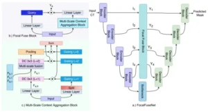
A Reverse Mamba Attention Network for Pathological Liver Segmentation
Authors: Jun Zeng, Debesh Jha, Ertugrul Aktas, Elif Keles, Alpay Medetalibeyoglu, Matthew Antalek, Robert Lewandowski, Daniela Ladner, Amir A. Borhani, Gorkem Durak, Ulas Bagci
IEEE ICECCME 2025
Publication Year: 2025

OUTCOMES OF PANCREAS CANCER DETECTED DURING PANCREAS CYST SURVEILLANCE IS SIMILAR TO INCIDENT CANCER
Authors: Andrea Mia Bejar
DDW 2025
Publication Year: 2025
RISK FACTORS FOR DEVELOPMENT OF PANCREAS ADENOCARCINOMA AFTER INITIAL DIAGNOSIS OF INCIDENTAL PANCREAS CYST
Authors: Andrea Mia Bejar et al
DDW 2025
Publication Year: 2025
A Conceptual Framework for Applying Ethical Principles of AI to Medical Practice
Authors: Debesh Jha et al
Bioengineering
Publication Year: 2025

Liver Cirrhosis Stage Estimation from MRI with Deep Learning
Authors: Jun Zeng, Debesh Jha, Ertugrul Aktas, Elif Keles, Alpay Medetalibeyoglu, Matthew Antalek, Federica Proietto Salanitri, Amir Borhani, Daniela Ladner, Gorkem Durak, Ulas Bagci
IEEE EMBC 2025
Publication Year: 2025


Conditional Visuo-Textual Prompt Learning for Medical Image Analysis
Authors: Raffaele Mineo et al
IEEE ISBI 2025
Publication Year: 2025
Is Long Range Sequential Modeling Necessary for Colorectal Tumor Segmentation?
Authors: Abhishek Srivastava, et al.
IEEE ISBI 2025
Publication Year: 2025

Adaptive Aggregation Weights for Federated Segmentation of Pancreas MRI
Authors: Hongyi Pan, Gorkem Durak, Zheyuan Zhang, Yavuz Taktak, Elif Keles, Halil Ertugrul Aktas, Alpay Medetalibeyoglu, Yury Velichko, Concetto Spampinato, Ivo Schoots, Marco J Bruno, Rajesh N Keswani, Pallavi Tiwari, Candice Bolan, Tamas Gonda, Michael G Goggins, Michael B Wallace, Ziyue Xu, Ulas Bagci
IEEE ISBI 2025
Publication Year: 2025

IPMN Risk Assessment under Federated Learning Paradigm
Authors: Hongyi Pan, Ziliang Hong, Gorkem Durak, Elif Keles, Halil Ertugrul Aktas, Yavuz Taktak, Alpay Medetalibeyoglu, Zheyuan Zhang, Yury Velichko, Concetto Spampinato, Ivo Schoots, Marco J Bruno, Pallavi Tiwari, Candice Bolan, Tamas Gonda, Frank Miller, Rajesh N Keswani, Michael B Wallace, Ziyue Xu, Ulas Bagci
IEEE ISBI 2025
Publication Year: 2025
A New Logic For Pediatric Brain Tumor Segmentation
Authors: Max Bengtsson, Elif Keles, Gorkem Durak, Syed Anwar, Yuri S Velichko, Marius G Linguraru, Angela J Waanders, Ulas Bagci
IEEE ISBI 2025
Publication Year: 2025


Long-Term Follow-up of Patients Recovering From Moderate-To-Severe COVID-19
Authors: Gorkem Durak et al
INTERNATIONAL CONFERENCE AMERICAN THORACIC SOCIETY (ATS) 2025
Publication Year: 2025
Radiomic Features Detect Interstitial Lung Disease in Patients with Systemic Sclerosis
Authors: Alec Peltekian et al
International Conference American Thoracic Society (ATS) 2025
Publication Year: 2025
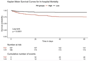
Predictive strength of inflammatory scores for in-hospital mortality in infective endocarditis
Authors: Vedat Cicek, Almina Erdem, Sahhan Kilic, Burak Tay, Mustafa Kamil Yemis, Solen Taslicukur, Mustafa Oguz, Ahmet Oz, Murat Selcuk, Tufan Cinar, Ulas Bagci
Herz
Publication Year: 2025

A New Risk Prediction Model for the Assessment of Myocardial Injury in Elderly Patients Undergoing Non-Elective Surgery
Authors: Cicek, V., Babaoglu, M., Saylik, F., Yavuz, S., Mazlum, A. F., Genc, M. S., Altinisik, H., Oguz, M., Korucu, B. C., Hayiroglu, M. I., Cinar, T., & Bagci, U.
Journal of Cardiovascular Development and Disease (2025)
Publication Year: 2025

A New Model for Prediction of Myocardial Injury of Non-Elective Surgery in Elderly
Authors: Vedat Cicek et al
ACC (2025)
Publication Year: 2025


Towards Synergistic Deep Learning Models for Volumetric Cirrhotic Liver Segmentation in MRIs
Authors: Vandan Gorade, Onkar Susladkar, Gorkem Durak, Elif Keles, Ertugrul Aktas, Timurhan Cebeci, Alpay Medetalibeyoglu, Daniela P. Ladner, Debesh Jha, and Ulas Bagci
SPIE Medical Imaging 2025
Publication Year: 2025


MDNet: Multi-Decoder Network for Abdominal CT Organs Segmentation
Authors: Debesh Jha, Nikhil Tomar, Koushik Biswas, Gorkem Durak, Matthew Antalek, Zheyuan Zhang, Bin Wang, Md Mostafijur Rahman, Hongyi Pan, Alpay Medetalibeyoglu, Abhijit Das, Yury Velichko, Daniela P. Ladner, Amir A. Borhani, Ulas Bagci
IEEE ICASSP 2025
Publication Year: 2025


Frequency-Based Federated Domain Generalization for Polyp Segmentation
Authors: Hongyi Pan, Debesh Jha, Koushik Biswas, Ulas Bagci
IEEE ICASSP 2025
Publication Year: 2025


Transformer-Enhanced Iterative Feedback Mechanism for Polyp Segmentation
Authors: Nikhil Tomar, Debesh Jha, Koushik Biswas, Ulas Bagci
IEEE ICASSP 2025
Publication Year: 2025

Uncertainty-Guided Coarse-to-Fine Tumor Segmentation with Anatomy-Aware Post-Processing
Authors: Ilkin Sevgi Isler, David Mohaisen, Curtis Lisle, Damla Turgut, Ulas Bagci
IEEE ACDSA 2025
Publication Year: 2025

Bridging the Gap in Pancreatic Imaging: A New Era of AI-Powered Precision
Authors: Gorkem Durak and Ulas Bagci
Journal of the Pancreas - 2025
Publication Year: 2025


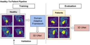
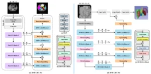
Healthy-to-Patients Deep learning for Automated Left Atrial Segmentation and Function analysis from Short-Axis Cine CMR
Authors: M. Elbayumi et al.
SCMR 2025
Publication Year: 2025
HCC Detection in the Wild: AI models for automated MRI HCC detection.
Authors: Matthew Antalek, Robert Lewandowski, and Ulas Bagci
SIR Annual Scientific Meeting - 2025
Publication Year: 2025
Large-scale multi-center CT and MRI segmentation of pancreas with deep learning (PanSegNet)
Authors: Zheyuan Zhang, Elif Keles, Gorkem Durak, Yavuz Taktak, Onkar Susladkar, Vandan Gorade, Debesh Jha, Asli C. Ormeci, Alpay Medetalibeyoglu, Lanhong Yao, Bin Wang, Ilkin Sevgi Isler, Linkai Peng, Hongyi Pan, Camila Lopes Vendrami, Amir Bourhani, Yury Velichko, Boqing Gong, Concetto Spampinato, Ayis Pyrros, Pallavi Tiwari, Derk C.F. Klatte, Megan Engels, Sanne Hoogenboom, Candice W. Bolan, Emil Agarunov, Nassier Harfouch, Chenchan Huang, Marco J. Bruno, Ivo Schoots, Rajesh N. Keswani, Frank H. Miller, Tamas Gonda, Cemal Yazici, Temel Tirkes, Baris Turkbey, Michael B. Wallace, Ulas Bagci
MEDICAL IMAGE ANALYSIS - 2025
Publication Year: 2025

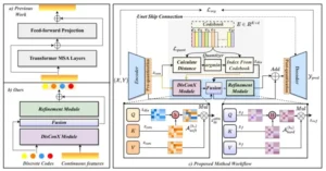
DiffBoost: Enhancing Medical Image Segmentation via Text-Guided Diffusion Model
Authors: Zheyuan Zhang, Lanhong Yao, Bin Wang, Debesh Jha, Gorkem Durak, Elif Keles, Alpay Medetalibeyoglu, Ulas Bagci
IEEE Transactions on Medical Imaging
Publication Year: 2025
ProFONet: Prototypical Feature Space Optimized Network for Few Shot Classification
Authors: Abhijit Das, Vandan Gorade, Debesh Jha, Koushik Biswas, Pethuru Raj, Ulas Bagci
ICPR 2024
Publication Year: 2024

SynergyNet: Bridging the Gap Between Discrete and Continuous Representations for Precise Medical Image Segmentation.
Authors: Gorade, Vandan, et al.
WACV 2024
Publication Year: 2024

Beyond Self-Attention: Deformable Large Kernel Attention for Medical Image Segmentation.
Authors: Azad, Reza, et al.
WACV 2024
Publication Year: 2024

Estimation of Acoustic Emission Arrival Time in Concrete Structures Using Convolutional Neural Network
Authors: Omair Inderyas, Ninel Alver, Aydin Kaya, Ulas Bagci
Proceedings of the 42nd IMAC (2024)
Publication Year: 2024


Foundational AI Models and Modern Medical Practice
Authors: Alpay Medetalibeyoglu, Yury Velichko, Eric Hart, MD, Ulas Bagci.
British Journal of Radiology AI
Publication Year: 2024


Variational Quantum Neural Network for Modeling and Solving Heat and Mass Transfer Problems Occurring in Electric Contact Phenomena
Authors: Merey M. Sarsengeldin; Zeeshan Ahmad; Ulas Bagci
IEEE 69th Holm Conference on Electrical Contacts (HOLM) (2024)
Publication Year: 2024

𝗔𝗱𝘃𝗮𝗻𝗰𝗲𝘀 𝗳𝗼𝗿 𝗠𝗮𝗻𝗮𝗴𝗶𝗻𝗴 𝗣𝗮𝗻𝗰𝗿𝗲𝗮𝘁𝗶𝗰 𝗖𝘆𝘀𝘁𝗶𝗰 𝗟𝗲𝘀𝗶𝗼𝗻𝘀: 𝗜𝗻𝘁𝗲𝗴𝗿𝗮𝘁𝗶𝗻𝗴 𝗜𝗺𝗮𝗴𝗶𝗻𝗴 𝗮𝗻𝗱 𝗔𝗜 𝗜𝗻𝗻𝗼𝘃𝗮𝘁𝗶𝗼𝗻𝘀
Authors: Deniz Seyithanoglu, Gorkem Durak, Elif Keles, Alpay Medetalibeyoglu, Ziliang Hong, Zheyuan Zhang, Yavuz B. Taktak, Timurhan Cebeci, Pallavi Tiwari, Yuri S. Velichko, Cemal Yazici, Temel Tirkes, Frank H. Miller, Rajesh N. Keswani, Concetto Spampinato, Michael B. Wallace and Ulas Bagci
Cancers (2024)
Publication Year: 2024

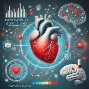
Predictive Value of Inflammatory Scores for Left Atrium Thrombosis in Ischemic Stroke Without Atrial Fibrillation
Authors: Vedat Cicek et al.
Medicina - 2024
Publication Year: 2024
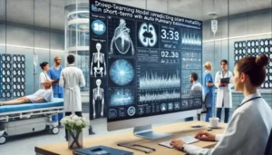
Predicting Short-Term Mortality in Patients With Acute Pulmonary Embolism With Deep Learning
Authors: Vedat Cicek et al.,
CIRCULATION JOURNAL
Publication Year: 2024

Mortality Prediction of Pulmonary Embolism Patients with Deep Learning and XGBoost
Authors: Vedat Cicek et al.
IEEE ICECCME 2024
Publication Year: 2024
Machine learning for prognostic prediction in coronary artery disease with SPECT data: a systematic review and meta-analysis
Authors: Vedat Cicek, et al.
EJNMMI Research
Publication Year: 2024

Evidential Federated Learning for Skin Lesion Image Classification
Authors: Rutger Hendrix et al
ICPR 2024
Publication Year: 2024
AI-powered contrast-free cardiovascular magnetic resonance imaging for myocardial infarction
Authors: Vedat Cicek and Ulas Bagci
FRONTIERS CARDIOVASCULAR MEDICINE
Publication Year: 2024

CT Liver Segmentation via PVT-based Encoding and Refined Decoding
Authors: Jha, Debesh, et al.
IEEE ISBI 2024 (Oral)
Publication Year: 2024
Deep learning based automated pancreas segmentation and volumetry measurement in patients with acute pancreatitis
Authors: Elif Keles, Gorkem Durak, Ziliang Hong, Jasson Barrios, Paya Sarraf,Matthew Antalek, Zheuyan Zhang, Brian Boulay, Ashley Vareedayah, Brian Layden,Temel Tirkes, Cemal Yazici, Ulas Bagci
APA/JPS/CAP/IAP 2024 Annual Meeting
Publication Year: 2024

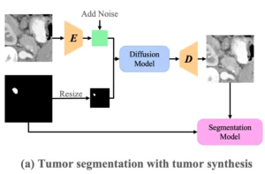
Optimizing Synthetic Data for Enhanced Pancreatic Tumor Segmentation
Authors: Linkai Peng, Zheyuan Zhang, Gorkem Durak, Frank H. Miller, Alpay Medetalibeyoglu, Michael B. Wallace, Ulas Bagci
MICCAI AIPAD 2024
Publication Year: 2024

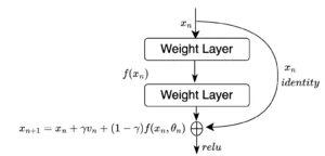
Novel Momentum-Based Deep Learning Techniques for Medical Image Classification and Segmentation
Authors: AKoushik Biswas, Ridal Pal, Shaswat Patel, Debesh Jha, Meghana Karri, Amit Reza, Gorkem Durak, Alpay Medetalibeyoglu, Matthew Antalek, Yury Velichko, Daniela Ladner, Amir Borhani, Ulas Bagci
MICCAI MLMI 2024
Publication Year: 2024
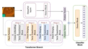
Classification of Endoscopy and Video Capsule Images using Hybrid Model
Authors: Aliza Subedi, Smriti Regmi, Nisha Regmi, Bhumi Bhusal, Ulas Bagci, Debesh Jha
MICCAI CaPTion 2024
Publication Year: 2024

Practical and Ethical Considerations for Generative AI in Medical Imaging
Authors: Debesh Jha, Ashish Rauniyar, Desta Haileselassie Hagos, Vanshali Sharma,Nikhil Kumar Tomar, Zheyuan Zhang, Ilkin Isler, Gorkem Durak, Michael Wallace, Cemal Yazici, Tyler Berzin, Koushik Biswas, Ulas Bagci
MICCAI EPIMI 2024
Publication Year: 2024

AI-ADC: Channel and Spatial Attention-Based Contrastive Learning to Generate ADC Maps from T2W MRI for Prostate Cancer Detection
Authors: Ozyoruk et al.
Journal of Personalized Medicine
Publication Year: 2024
Advancing Pediatric Brain Tumor Diagnosis: A Deep Learning Approach to Multiclass Segmentation in CBTN data: Feasibility Study
Authors: Max Bengtsson, et al
CBTN (Children Brain Tumor Network) Summit 2024
Publication Year: 2024

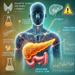
The association of volumetric changes and disease severity with endocrine dysfunction following acute pancreatitis
Authors: Elif Keles, Gorkem Durak,Jasson Barrios, Ziliang Hong, Paya Sarraf,Matthew Antalek, Zheuyan Zhang, Brian Boulay, Ashley Vareedayah, Brian Layden,Temel Tirkes, Ulas Bagci,Cemal Yazici,
APA/JPS/CAP/IAP 2024 Annual Meeting
Publication Year: 2024
Patients with acute pancreatitis have significant decline in pancreas volume over time
Authors: Jasson Barrios, Elif Keles, Gorkem Durak, Ziliang Hong, Paya Sarraf,Matthew Antalek, Zheuyan Zhang, Brian Boulay, Ashley Vareedayah, Brian Layden,Temel Tirkes,Ulas Bagci,Cemal Yazici
APA/JPS/CAP/IAP 2024 Annual Meeting
Publication Year: 2024

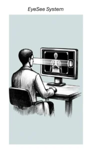
EyeSee: Integrating Deep Learning for ImageAnalysis
Authors: Bin Wang and Ulas Bagci
Medical Image Perception Society (MIPS) XX 2024.
Publication Year: 2024
The Role of CO-RADS Scoring System in the Diagnosis of COVID-19 Infection and its Correlation with Clinical Signs
Authors: Şenol Çomoğlu, Sinan Öztürk, Ahmet Topçu, Fatma Kulalı, Aydın Kant, Resul Sobay, Mustafa Arslan, Hanife Şeyda Ülgür, Uğur Kostakoğlu, Eyüp Veli Küçük, Hanife Nur Karakoç, Merve Çağlar, Gülsüm Uzuğ, Ulas Bagci , Ömer Faruk Özkan, Gürdal Yılmaz
Curr Med Imaging - 2022
Publication Year: 2022

Early stage lung cancer detection from speech sounds in natural environments.
Authors: Haydar Ankishan, et al.
Biomedical Signal Processing and Control
Publication Year: 2024

Deformable Capsules: A Novel Capsule Network Architecture for Object Detection.”
Authors: Naji Khosravan, Rodney LaLonde, Ulas Bagci
Advanced Intelligent Systems (Willey)
Publication Year: 2024
Utilizing Chest Radiographs (CXRS) for Kidney Failure Prognostication and Early Risk Stratification with Prospective and External validation
Authors: Theo Dapamede et al.
RSNA 2024
Publication Year: 2024
MRI Intensity Standardization via Prototype Learning fro Improved Oncological Analysis of Heterogenous Datasets
Authors: Meghana Karri, et al.
RSNA 2024
Publication Year: 2024
Limited Utility of RECIST 1.1 in Predicting Pathologic Response to Neoadjuvant Radiotherapy in Myxoid Liposarcoma
Authors: Yuri Velichko et al.
RSNA 2024
Publication Year: 2024
Increased Tumor Size on MRI Predicts Pathological Response to Neoadjuvant Radiotherapy in Undifferentiated Pleomorphic Sarcoma
Authors: Yuri Velichko et al.
RSNA 2024
Publication Year: 2024
EyeSee: Real Radiology Room Experience with Eye Tracking and Deep Learning
Authors: Bin Wang, et al.
RSNA 2024
Publication Year: 2024
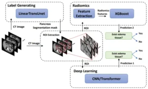
Detection of Peri-Pancreatic Edema Using Deep Learning and Radiomics
Authors: ZIliang Hong, et al.
RSNA 2024
Publication Year: 2024
Enhanced Prediction of Lung Cancer Risk from Imaging Biomarkers in Chests Radiographs: An External Validation Study
Authors: Theo Dapamede et al.
RSNA 2024
Publication Year: 2024
Imaging Assessment of Response in Soft Tissue Sarcoma After Neoadjuvant Radiotheraphy
Authors: Yuri Velichko et al.
RSNA 2024
Publication Year: 2024

Domain Generalization with Correlated Style Uncertainty.
Authors: Zhang, Zheyuan, et al.
WACV 2024
Publication Year: 2024
INCODE: Implicit Neural Conditioning With Prior Knowledge Embeddings
Authors: Kazerouni, Amirhossein, et al.
WACV 2024
Publication Year: 2024

GazeGNN: A Gaze-Guided Graph Neural Network for Chest X-Ray Classification.
Authors: Wang, Bin, et al.
WACV 2024
Publication Year: 2024
Harmonized Spatial and Spectral Learning for Robust and Generalized Medical Image Segmentation
Authors: Gorade, Vandan, et al.
ICPR 2024
Publication Year: 2024

Domain Generalization with Fourier Transform and Soft Thresholding.
Authors: Pan, Hongyi, et al.
IEEE International Conference on Acoustics, Speech and Signal Processing (ICASSP) 2024
Publication Year: 2024
Explainable Transformer Prototypes for Medical Diagnoses.
Authors: Demir, Ugur, et al.
IEEE ISBI 2024
Publication Year: 2024

THE BOSTON ERCP DATASET: A VIDEO DATASET FOR ADVANCED ENDOSCOPY
Authors: Mark E. Geissler, et al.
DDW 2024
Publication Year: 2024


Deep Learning-Based Detection and Classification of Bone Lesions on Staging Computed Tomography in Prostate Cancer: A Development Study
Authors: Belue, Mason J., et al.
Academic Radiology - 2024
Publication Year: 2024
SAM-EG: Segment Anything Model with Egde Guidance framework for efficient Polyp Segmentation
Authors: Quoc-Huy Trinh, Hai-Dang Nguyen, Bao-Tram Nguyen Ngoc, Debesh Jha, Ulas Bagci, Minh-Triet Tran
BMVC 2024
Publication Year: 2024

PGDS: Pose-Guidance Deep Supervision for Mitigating Clothes-Changing in Person Re-Identification
Authors: Quoc-Huy Trinh, Nhat-Tan Bui, Dinh-Hieu Hoang, Phuoc-Thao Vo Thi, Hai-Dang Nguyen, Debesh Jha, Ulas Bagci, Ngan Le, Minh-Triet Tran
IEEE AVSS 2024
Publication Year: 2024
Predicting Short Term Mortality In Patients With Acute Pulmonary Embolism With Deep Learning
Authors: V Cicek, et al.
ICNC-CT 2024
Publication Year: 2024

Evaluation of pan-Immuno-Inflammation value for In-hospital mortality in acute pulmonary embolism patients
Authors: Çiçek, Vedat, et al.
Revista de Investigacion Clinica -2024
Publication Year: 2024

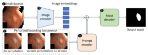
PP-SAM: Perturbed Prompts for Robust Adaption of Segment Anything Model for Polyp Segmentation
Authors: Md M. Rahman, M. Munir, D. Jha, U. Bagci, R. Marculescu
CVPR DEF-AI-MIA 2024
Publication Year: 2024

AI-Powered Road Network Prediction with Multi-Modal Data
Authors: Necip Gengec, Engin Tari, Ulas Bagci
Earth Science Informatics - 2024
Publication Year: 2024

Methods of artificial intelligence-assisted infrastructure assessment using mixed reality systems.
Authors: Karaaslan, Enes, et al.
US Patent - 2024
Publication Year: 2024


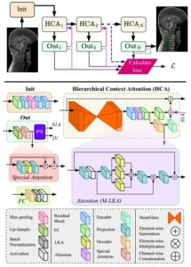
HCA-Net: Hierarchical Context Attention Network for Intervertebral Disc Semantic Labeling
Authors: Bozorgpour, Afshin, et al.
IEEE ISBI 2024
Publication Year: 2024
Federated Learning for Medical Applications: A Taxonomy, Current Trends, Challenges, and Future Research Directions
Authors: Rauniyar, Ashish, et al.
IEEE Internet of Things Journal
Publication Year: 2024


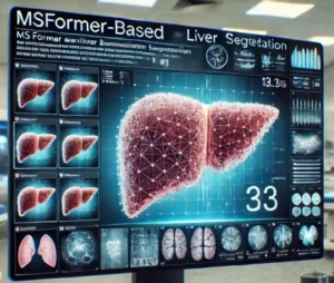
Enhancing Liver Segmentation Outcomes with MSFormer-based AI System
Authors: Jha, Debesh, et al.
DDW 2024
Publication Year: 2024
Adaptive Smooth Activation for Improved Disease Diagnosis and Organ Segmentation from Radiology Scans
Authors: Koushik Biswas, Debesh Jha, Nikhil Kumar Tomar, Gorkem Durak, Alpay Medetalibeyoglu, Matthew Antalek, Yury Velichko, Daniela Ladner, Amir Bohrani, Ulas Bagci
MICCAI 2024
Publication Year: 2024

Rethinking Intermediate Layers design in Knowledge Distillation for Kidney and Liver Tumor Segmentation
Authors: Gorade, Vandan, et al.
IEEE ISBI 2024
Publication Year: 2024

Enhancing Colonoscopy Outcomes with DAPODET-based AI for Real-time sessile serrated Polyp Detection
Authors: Das, Abhijit, et al.
DDW 2024
Publication Year: 2024

Healthy-to-Patients Domain-Adaptive Deep Learning for Time-Resolved Segmentation of Left Atrium in Short-Axis Cine MRI Images
Authors: M. Elbayumi, et al.
ISMRM 2024
Publication Year: 2024

PAM-UNet: Shifting Attention on Region of Interest in Medical Images
Authors: A. Das, D. Jha, V. Gorade, K. Biswas, H. Pan, Z. Zhang, D. P. Ladner, Y. Velichko, A. Borhani, and U. Bagci
IEEE EBMC 2024
Publication Year: 2024
Prediction of MRI-Induced Power Absorption in Patients with DBS Leads
Authors: Yalcin Tur, et al.
IEEE EMBS 2024
Publication Year: 2024

FuseNet: Self-Supervised Dual-Path Network for Medical Image Segmentation
Authors: Kazerouni, Amirhossein, et al.
IEEE ISBI 2024
Publication Year: 2024
Leveraging Unlabeled Data for 3D Medical Image Segmentation through Self-Supervised Contrastive Learning
Authors: Karimijafarbigloo, Sanaz, et al.
IEEE ISBI 2024
Publication Year: 2024
A review of prognostic prediction of coronary artery disease patients with myocardial perfusion scintigraphy and artificial intelligence
Authors: V. Cicek, et al.
ICNC-CT 2024
Publication Year: 2024

ControlPolypNet: Towards Controlled Colon Polyp Synthesis for Improved Polyp Segmentation
Authors: V. Sharma, A. Kumar, D. Jha, M.K. Bhuyan, P. Das, U. Bagci.
CVPR DCAMI 2024
Publication Year: 2024
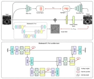
A Probabilistic Hadamard U-Net for MRI Bias Field Correction
Authors: Zhu, Xin, et al.
MICCAI MLMI 2024
Publication Year: 2024
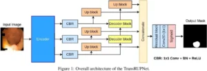
TransRUPNet for Improved Out-of-Distribution Generalization in Polyp Segmentation
Authors: D. Jha, N. K. Tomar, D. Bhattacharya, K. Biswas, U. Bagci
IEEE EBMC 2024
Publication Year: 2024

Detection of Peri-Pancreatic Edema using Deep Learning and Radiomics Techniques
Authors: Z. Hong, D. Jha, K. Biswas, Z. Zhang, Y. Velichko, C. Yazici, T. Tirkes, A. Borhani, B. Turkbey, A. Medetalibeyoglu, G. Durak, U. Bagci
IEEE EBMC 2024
Publication Year: 2024
Multichannel Orthogonal Transform-Based Perceptron Layers for Efficient ResNets
Authors: H. Pan, E. Hamdan, X. Zhu, S. Atici and A. E. Cetin
IEEE Transactions on Neural Networks and Learning Systems
Publication Year: 2024

COVID-19 Detection from Respiratory Sounds with Hierarchical Spectrogram Transformers
Authors: Aytekin, Idil, et al.
IEEE JBHI - 2024
Publication Year: 2024

TransNetR: Transformer-based Residual Network for Polyp Segmentation with Multi-Center Out-of-Distribution Testing
Authors: Debesh Jha, Nikhil Kumar Tomar, Vanshali Sharma, Ulas Bagci
MIDL 2023
Publication Year: 2023
The past, current, and future of neonatal intensive care units with artificial intelligence: a systematic review
Authors: Keles, Elif, and Ulas Bagci
npj Digital Medicine - 2023
Publication Year: 2023


Ensemble Learning with Residual Transformer for Brain Tumor Segmentation
Authors: Yao, Lanhong, et al.
IEEE ISBI 2023
Publication Year: 2023
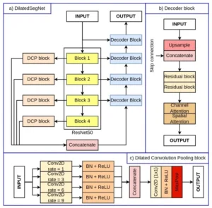
DilatedSegNet: A Deep Dilated Segmentation Network for Polyp Segmentation
Authors: Tomar, NK., Jha, D., Bagci, U.
SPIE Medical Imaging 2023
Publication Year: 2023

Artificial Intelligence and Infectious Disease Imaging
Authors: Chu, Winston T., et al.
The Journal of Infectious Diseases - 2023
Publication Year: 2023

A review of deep learning and radiomics approaches for pancreatic cancer diagnosis from medical imaging
Authors: Yao, Lanhong; Zhang, Zheyuan; Keles, Elif; Yazici, Cemal; Tirkes, Temel; Bagci, Ulas
Current Opinion in Gastroenterology
Publication Year: 2023


TransResU-Net: Transformer based ResU-Net for Real-Time Colonoscopy Polyp Segmentation
Authors: Tomar, Nikhil Kumar, et al.
IEEE EMBC 2023
Publication Year: 2023
Monkeypox Diagnosis with Interpretable Deep Learning
Authors: Ahsan, Md. Manjurul, Ali, Md. Shahin, Hassan, Md. Mehedi, Abdullah, Tareque Abu, Gupta, Kishor Datta, Bagci, Ulas, Kaushal, Chetna, Soliman, Naglaa F.
IEEE Access - 2023
Publication Year: 2023


Radiomics Boosts Deep Learning Model for IPMN Classification
Authors: Yao L, Zhang Z, Demir U, Keles E, Vendrami C, Agarunov E, Bolan C, Schoots I, Bruno M, Keswani R, Miller F, Gonda T, Yazici C, Tirkes T, Wallace M, Spampinato C, Bagci U.
MICCAI MLMI 2023
Publication Year: 2023

A Fully Automatic AI System for Pancreas Segmentation from Multicenter MRI Scans
Authors: Zheyuan Zhang, et al.
DDW 2023
Publication Year: 2023

RUPNet: residual upsampling network for real-time polyp segmentation
Authors: Tomar, Nikhil Kumar, Ulas Bagci, and Debesh Jha.
SPIE Medical Imaging 2023
Publication Year: 2023

Laplacian-Former: Overcoming the Limitations of Vision Transformers in Local Texture Detection
Authors: Azad, Reza, et al
MICCAI 2023
Publication Year: 2023

Self-supervised Semantic Segmentation: Consistency over Transformation
Authors: S Karimijafarbigloo, R Azad, et al.
ICCV 2023
Publication Year: 2023


Gastrointestinal Disease Diagnosis with Hybrid Model of Capsules and CNNs
Authors: Sarsengeldin, Merey, et al.
IEEE EIT - 2023
Publication Year: 2023
Deep Learning Algorithms for Pancreas Segmentation from Radiology Scans: A Review
Authors: Zhang, Zheyuan, et al.
Advances in Clinical Radiology - 2023
Publication Year: 2023

GastroVision: A Multi-class Endoscopy Image Dataset for Computer Aided Gastrointestinal Disease Detection
Authors: Jha, Debesh, et al.
ICML Workshop 2023
Publication Year: 2023

AI in Clinical Medicine: A Practical Guide for Healthcare Professionals
Authors: Michael F. Byrne (Editor), Nasim Parsa (Co-Editor), Alexandra T. Greenhill (Co-Editor), Daljeet Chahal (Co-Editor), Omer Ahmad (Co-Editor), Ulas Bagci (Co-Editor).
WILEY - 2023
Publication Year: 2023
Multi-Institutional Large-Scale Validation of 8 Methods for Automatic Knee MRI Segmentation for Use in Clinical Trials
Authors: Dam, E. B., et al.
Osteoarthritis and Cartilage 2022
Publication Year: 2022
Real-time multi-class helmet violation detection using few-shot data sampling technique and yolov8
Authors: Aboah, Armstrong, et al.
CVPRW 2023
Publication Year: 2023

Deepsegmenter: Temporal action localization for detecting anomalies in untrimmed naturalistic driving videos
Authors: Aboah, Armstrong, et al.
CVPRW 2023
Publication Year: 2023

An Efficient Multi-Scale Fusion Network for 3D Organ at Risk (OAR) Segmentation
Authors: Srivastava, Abhishek, et al.
IEEE EMBC 2023
Publication Year: 2023

Self-Supervised Learning for Organs At Risk and Tumor Segmentation with Uncertainty Quantification
Authors: Ilkin Isler, Debesh Jha, Curtis Lisle, et al.
IEEE ICECCME 2023
Publication Year: 2023
GazeSAM: Interactive Image Segmentation with Eye Gaze and Segment Anything Model
Authors: Wang, Bin, et al.
NeurIPS Workshop 2023
Publication Year: 2023
Relational reasoning network for anatomical landmarking
Authors: Torosdagli, Neslisah, et al.
Journal of Medical Imaging - 2023
Publication Year: 2023
AI Empowered Automatic Volume Delineation of Liver from CT Scans for Diagnostic Workflow
Authors: Ugur Demir, Zheyuan Zhang, Bin Wang, et al.
DDW 2023
Publication Year: 2023
Deep Learning Models in the Classification of Intraductal Papillary Mucinous Neoplasms with Visual Explanation Methods to Highlight Areas of Interest for Algorithm Decision Process
Authors: Engels, M. M., et al.
Gastroenterology 2022
Publication Year: 2022
Enhancing organ at risk segmentation with improved deep neural networks
Authors: Isler, Ilkin, et al.
SPIE Medical Imaging 2022
Publication Year: 2022
Transformer-based Generative Adversarial Network for Liver Segmentation
Authors: Demir, Ugur, et al.
International Conference on Image Analysis and Processing (ICIAP) 2022
Publication Year: 2022
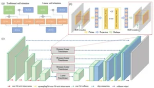
Dynamic Linear Transformer for 3D Biomedical Image Segmentation
Authors: Zhang, Z., & Bagci, U.
MICCAI MLMI
Publication Year: 2022
Out of Distribution Detection, Generalization, and Robustness Triangle with Maximum Probability Theorem
Authors: Marvasti, Amir Emad, et al.
IEEE ICECCME 2022
Publication Year: 2022

Automatic Polyp Segmentation with Multiple Kernel Dilated Convolution Network
Authors: Tomar, Nikhil Kumar, et al.
IEEE 35th International Symposium on Computer-Based Medical Systems (CBMS)
Publication Year: 2022

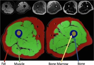
Musculoskeletal MR Image Segmentation with Artificial Intelligence
Authors: Keles, E., Irmakci, I., Bagci, U.
Advances in Clinical Radiology
Publication Year: 2022
Detecting COVID-19 from respiratory sound recordings with transformers
Authors: Aytekin, Idil, et al.
SPIE Medical Imaging 2022
Publication Year: 2022

Multi-Contrast MRI Segmentation Trained on Synthetic Images
Authors: Irmakci, I., Unel, Z. E., Ikizler-Cinbis, N., & Bagci, U.
IEEE EMBC 2022
Publication Year: 2022
No-Reference Image Quality Assessment Of T2-Weighted Magnetic Resonance Images In Prostate Cancer Patients.
Authors: Masoudi, S., Harmon, S., Mehralivand, S., Lay, N., Bagci, U., Wood, B.J., Pinto, P.A., Choyke, P. and Turkbey, B. (2021).
IEEE ISBI - 2021
Publication Year: 2021

Design and Rationale for the Use of Magnetic Resonance Imaging Biomarkers to Predict Diabetes After Acute Pancreatitis in the Diabetes Related to Acute Pancreatitis and Its Mechanisms Study: From the Type 1 Diabetes in Acute Pancreatitis Consortium
Authors: Tirkes T., et al,
Pancreas 2022
Publication Year: 2022
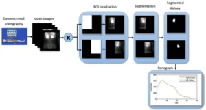
An automatic segmentation framework for computer-assisted renal scintigraphy
Authors: Rahimi, A., Hosntalab, M., Babapour, F., Amoui, M., Bagci, U.
Medical & Biological Engineering & Computing (Springer)
Publication Year: 2022

Neural Transformers for Intraductal Papillary Mucosal Neoplasms (IPMN) Classification in MRI images
Authors: Proietto Salanitri, F. et al.
IEEE EMBC 2022
Publication Year: 2022

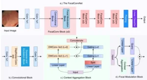
Video Capsule Endoscopy Classification using Focal Modulation Guided Convolutional Neural Network
Authors: Srivastava, Abhishek, et al.
IEEE 35th International Symposium on Computer-Based Medical Systems (CBMS) 2022
Publication Year: 2022

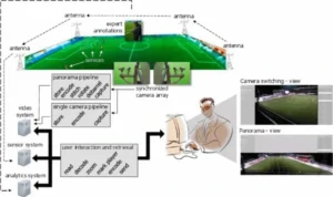
Video Analytics in Elite Soccer: A Distributed Computing Perspective
Authors: Jha, Debesh, et al.
IEEE 12th Sensor Array and Multichannel Signal Processing Workshop (SAM)
Publication Year: 2022

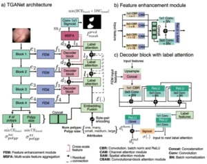
TGANet: Text-guided attention for improved polyp segmentation
Authors: Tomar, N. K., Jha, D., Bagci, U., & Ali, S.
MICCAI 2022
Publication Year: 2022

The Role of CO-RADS Scoring System in the Diagnosis of COVID-19 Infection and its Correlation with Clinical Signs
Authors: Çomoglu, S., Öztürk, S., Topçu, A., Kulali, F., Kant, A., Sobay, R., … & Yilmaz, G.
Current Medical Imaging - 2022
Publication Year: 2022
Hierarchical 3D Feature Learning for Pancreas Segmentation.
Authors: Proietto Salanitri, F., Bellitto, G., Irmakci, I., Palazzo, S., Bagci, U., & Spampinato, C.
MICCAI MLMI - 2021
Publication Year: 2021

Interpretable Deep Model for Predicting Gene-Addicted Non-Small-Cell Lung Cancer in CT Scans
Authors: Pino, Carmelo, Simone Palazzo, Francesca Trenta, et al.
IEEE ISBI
Publication Year: 2021
Information Bottleneck Attribution for Visual Explanations of Diagnosis and Prognosis
Authors: Demir, U., Irmakci, I., Keles, E., Topcu, A., Xu, Z., Spampinato, C., … & Bagci, U.
MICCAI MLMI - 2021
Publication Year: 2021
Missed Diagnosis of Pancreatic Ductal Adenocarcinoma Detection Using Deep Convolutional Neural Network.
Authors: Hoogenboom, S. A., Ravi, K., Engels, M. M., Irmakci, I., Keles, E., Bolan, C. W., … & Bagci, U
Gastroenterology - 2021
Publication Year: 2021
Deep Recurrent-Convolutional Model for Automated Segmentation of Craniomaxillofacial CT Scans.
Authors: Murabito, Francesca, Simone Palazzo, F. Proietto Salanitri, et al.
ICPR - 2021 (2020th conference published in 2021 due to covid)
Publication Year: 2021

Deep Multi-stage Model for Automated Landmarking of Craniomaxillofacial CT Scans
Authors: Palazzo, Simone, Giovanni Bellitto, Luca Prezzavento, et al.
IEEE ICPR -2021 (2020 conference was published in 2021 due to covid)
Publication Year: 2021
Quick Guide on Radiology Image Pre-processing for Deep Learning Applications in Prostate Cancer Research.
Authors: Masoudi, Samira, Stephanie AA Harmon, Sherif Mehralivand, et al.
Journal of Medical Imaging - 2021
Publication Year: 2021
Predicting RF Heating of Conductive Leads During Magnetic Resonance Imaging at 1.5 T: A Machine Learning Approach.
Authors: Zheng, C., Chen, X., Nguyen, B. T., et al.
IEEE EMBC 2021
Publication Year: 2021
The International Workshop on Osteoarthritis Imaging Knee MRI Segmentation Challenge: A Multi-Institute Evaluation and Analysis Framework on a Standardized Dataset
Authors: Desai, A. D., Caliva, F., Iriondo,
Radiology AI - 2021
Publication Year: 2021

Attention-guided Analysis of Infrastructure Damage with Semi-supervised Deep Learning.
Authors: Karaaslan, E., Bagci, U., & Catbas, F. N.
Automation in Construction - 2021
Publication Year: 2021


Morphometric and Functional Brain Connectivity Differentiates Chess Masters From Amateur Players
Authors: RaviPrakash, H., Anwar, S. M., Biassou, N. M., & Bagci, U.
Frontiers in Neuroscience - 2021
Publication Year: 2021

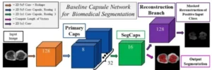
Capsules for Biomedical Image Segmentation
Authors: LaLonde, R., Xu, Z., Irmakci, I., Jain, S., & Bagci, U.
Medical image analysis - 2021
Publication Year: 2021

Maximum Probability Theorem: A Framework for Probabilistic Machine Learning
Authors: Marvasti, A. E., Marvasti, E. E., Bagci, U., & Foroosh, H
IEEE Transactions on Artificial Intelligence - 2021
Publication Year: 2021
A Novel Decision Support System for Long-Term Management of Bridge Networks
Authors: Karaaslan, E., Bagci, U., & Catbas, N
Applied Sciences, 11(13), 5928.
Publication Year: 2021
Deep Learning Based Staging of Bone Lesions from Computed Tomography Scans
Authors: Masoudi, S., Mehralivand, S., Harmon, S.A., et al.
IEEE Access - 2021
Publication Year: 2021
Machine Learning-Based Prediction of MRI-Induced Power Absorption in the Tissue in Patients With Simplified Deep Brain Stimulation Lead Models
Authors: Vu, J., Nguyen, B. T., Bhusal, B., Baraboo, J., Rosenow, J., Bagci, U., … & Golestanirad, L.
IEEE Transactions on Electromagnetic Compatibility - 2021
Publication Year: 2021

A Machine Learning-Based Prediction of the Micropapillary/Solid Growth Pattern in Invasive Lung Adenocarcinoma with Radiomics
Authors: He, B., Song, Y., Wang, L., et al.
Translational Lung Cancer Research - 2021
Publication Year: 2021

Brain Tumor Survival Prediction using Radiomics Features
Authors: Yousaf, S., Anwar, S, RaviPrakash, H., Bagci, U.
MICCAI RNO-AI 2020
Publication Year: 2020


Adipose Tissue Segmentation in Unlabeled Abdomen MRI Using Cross Modality Domain Adaptation
Authors: Masoudi, S., Razi, A., Gatlin, J., Turkbey, B., Bagci, U.
IEEE EMBC 2020
Publication Year: 2020

AI for the detection of COVID19 pneumonia on chest CT using multinational datasets.
Authors: Stephanie A. Harmon, Thomas H. Sanford, Sheng Xu, et al.
Nature Communications, 2020.
Publication Year: 2020
Variational Capsule Encoder
Authors: RaviPrakash, H., Anwar, SM., Bousquet, C., Bagci, U.
ICPR 2020
Publication Year: 2020
Instance-level Microtubule Tracking
Authors: Masoodi, S., Razi, A., Wright, C., Gatlin, J., Bagci, U.
IEEE Transactions on Medical Imaging - 2020
Publication Year: 2020
Diagnosing Colorectal Polyps in the Wild with Capsule Networks
Authors: LaLonde, R., Kandel, P., Spampinato, C., Wallace, M, Bagci, U.
IEEE ISBI 2020
Publication Year: 2020
Overall Survival Prediction in Gliomas Using Region-Specific Radiomic Features
Authors: Shaheen, A., Bagci, U., Mohy-ud-Din, H.
MICCAI RNO-AI - 2020
Publication Year: 2020
Integrating eye tracking and speech recognition accurately annotates MR brain images for deep learning: proof of principle
Authors: Stember, Joseph N., Haydar Celik, David Gutman, Nathaniel Swinburne, Robert Young, Sarah Eskreis-Winkler, Andrei Holodny, Sachin Jambawalikar , Bradford J Wood , Peter D Chang, Elizabeth Krupinski, Ulas Bagci
Radiology: Artificial Intelligence - 2020
Publication Year: 2020
Encoding High-Level Visual Attributes in Capsules for Explainable Medical Diagnoses
Authors: LaLonde, R., Torigian, D., Bagci, U.
MICCAI 2020
Publication Year: 2020
State-of-the-art in brain tumor segmentation and current challenges
Authors: Yousaf, Sobia, Harish RaviPrakash, Syed Muhammad Anwar, Nosheen Sohail, and Ulas Bagci.
MICCAI RNO-AI 2020
Publication Year: 2020
Semi-supervised deep learning for multi-tissue segmentation from multi-contrast MRI.
Authors: Anwar, Syed Muhammad, Ismail Irmakci, Drew A. Torigian, Sachin Jambawalikar, Georgios Z. Papadakis, Can Akgun, Jutta Ellermann, Mehmet Akcakaya, and Ulas Bagci.
Journal of Signal Processing Systems - 2020
Publication Year: 2020

Deep Convolutional Neural Networks Based Classification of Alzheimer’s Disease Using MRI Data
Authors: Nawaz, A., Anwar, S., Liaqat, R., Iqbal, J., Bagci, U.
IEEE INMIC 2020
Publication Year: 2020

The impact of COVID-19 on African American communities in the United States
Authors: Cyrus, Elena, Rachel Clarke, Dexter Hadley, Zoran Bursac, Mary Jo Trepka, Jessy G. Dévieux, Ulas Bagci
Health Equity - 2020
Publication Year: 2020

Deep Learning Provides Exceptional Accuracy to ECoG-based Functional Language Mapping for Epilepsy Surgery
Authors: RaviPrakash, H., Korostenskaja, M., Castillo, E., Lee, KH., Salinas, CM., Baumgartner, J., Scampinatoe, C., Bagci, U.
Frontiers in Neuroscience -
Publication Year: 2020

Analysis of Video Retinal Angiography with Deep Learning and Eulerian Magnification
Authors: Saha, S., LaLonde R., Carmack, A., Foroosh, H., Olson, J.C., Shaikh, S., Bagci, U.
Frontiers in Computer Science - 2020
Publication Year: 2020

AI in Gastroenterology. Current State of Play and Potential. How will it affect our practice and when?
Authors: Hoogenboom, S., Bagci, U., Wallace, MB.
Techniques in Gastrointestinal Endoscopy - 2020
Publication Year: 2020

Development of a Device-to-Image Registration Free Needle Guide for Magnetic Resonance Imaging-Guided Targeted Prostate Biopsy
Authors: Pankaj Kulkarni, Sakura Sikander, Pradipta Biswas, Sumit Laha, Heather Cornnell, Jeremy R Burt, Ulas Bagci, Sang-Eun Song.
Journal of Medical Devices - 2020
Publication Year: 2020

EEG based classification of long-term stress using psychological labeling
Authors: Saeed, Sanay Muhammad Umar, Syed Muhammad Anwar, Humaira Khalid, Muhammad Majid, and Ulas Bagci
Sensors - 2020
Publication Year: 2020

PET/CT and PET/MRI in Ophthalmic Oncology
Authors: Kalemaki MS, Karantanas AH, Exarchos D, Detorakis ET, Zoras O, Marias K, Millo C, Bagci U, Pallikaris I, Stratis A, Karatzanis I, Perisinakis K, Koutentakis P, Kontadakis GA, Spandidos DA, Tsatsakis A, Papadakis GZ.
International Journal of Oncology - 2020
Publication Year: 2020

Proceedings from the first global artificial intelligence in gastroenterology and endoscopy Summit
Authors: Parasa, S., Wallace, M., Bagci, U., Antonino, M., Berzin, T., Byrne, M., Celik, H., Farahani, K., Golding, M., Gross, S., and Jamali, V.
Gastrointestinal Endoscopy - 2020
Publication Year: 2020
Emotion Classification in Response to Tactile Enhanced Multimedia Using Frequency Domain Features of Brain Signals
Authors: Arsalan, A., Majid, M., Anwar, SM., Bagci, U.
IEEE EMBC 2019
Publication Year: 2019

Classification of Perceived Human Stress Using Psychological Signals
Authors: Arsalan, A., Majid, M., Anwar, SM., Bagci, U.
IEEE EMBC 2019
Publication Year: 2019
A Survey on Recent Advancements for AI-Enabled Radiomics in Neuro-Oncology
Authors: Anwar, S., Tooba, A., Rafique, K., RaviPrakash, H., Mohy-ud-din, H., Bagci, U.
MICCAI RNO-AI 2019
Publication Year: 2019

Deep Learning for Musculoskeletal Image Analysis
Authors: Irmakci, I., Anwar, S., Torigian, D., Bagci, U.
IEEE ASILOMAR 2019 (invited article)
Publication Year: 2019


Cross-modality Knowledge Transfer for Prostate Segmentation from CT Scans
Authors: Liu, Y., Khosravan, N., Liu, Y., Stember, J., Bagci, U., Jambawalikar, S.
MICCAI DART 2019
Publication Year: 2019

Weakly Supervised Segmentation by A Deep Geodesic Prior.
Authors: Mortazi, A., Khosravan, N., Torigian, DA., Kurugol, S., Bagci, U.
MICCAI MLMI 2019
Publication Year: 2019


PAN: Projective Adversarial Network for Medical Image Segmentation
Authors: Khosravan, N., Mortazi, A., Wallace, M., Bagci, U.
MICCAI 2019
Publication Year: 2019

INN: Inflated Neural Networks for IPMN Diagnosis
Authors: LaLonde, R., Tanner, I., Nikiforaki, K., Papadakis, GZ., Kandel, P., Bolan, CW., Wallace, M., Bagci, U.
MICCAI 2019
Publication Year: 2019
Lung and Pancreatic Tumor Characterization in the Deep Learning Era: Novel Supervised and Unsupervised Learning Approaches
Authors: Hussein, S., Kandel, MM., Bolan, CW., Wallace, MB., Bagci, U.
IEEE Transactions on Medical Imaging - 2019
Publication Year: 2019
A Collaborative Computer Aided Diagnosis (C-CAD) System with Eye Tracking, Sparse Attentional Model, and Deep Learning
Authors: Khosravan, N., Celik, H., Turkbey, B., Jones, E., Wood, B., Bagci, U.
Medical Image Analysis - 2019
Publication Year: 2019
Deep Geodesic Learning for Segmentation and Anatomical Landmarking
Authors: Neslisah Torosdagli, Denise K. Liberton, Payal Verma, Murat Sincan, Janice S. Lee, Ulas Bagci
IEEE Transactions on Medical Imaging - 2019
Publication Year: 2019
Artificial Intelligence Assisted Infrastructure Assessment Using Mixed Reality Systems
Authors: Karaaslan, E., Bagci, U., and Catbas, F.N.
Journal of Transportation Research - 2019
Publication Year: 2019

Eye-Tracking for Deep Learning Segmentation Using Convolutional Neural Networks
Authors: Stember, JN., … , Jambawalikar, S., Bagci, U.
Journal of Imaging Informatics in Medicine- 2019
Publication Year: 2019

18F-NaF PET/CT imaging in fibrous dysplasia of bone: implications for evaluation of disease activity and treatment
Authors: Papadakis, GZ., … , and Boyce, AM.
Journal of Bone and Mineral Research - 2019
Publication Year: 2019


Deep Learning to Classify Intraductal Papillary Mucinous Neoplasms Using Magnetic Resonance Imaging
Authors: Hurtado, S. Hussein, … , and M. Wallace.
Pancreas - 2019
Publication Year: 2019

Quality Assurance of Computer-Aided Detection and Diagnosis in Colonoscopy
Authors: Vinsard, DG., … , Wallace, MB.
Gastrointestinal Endoscopy - 2019
Publication Year: 2019
RETOUCH-The Retinal OCT Fluid Detection and Segmentation Benchmark and Challenge
Authors: Bugunovic, H., … , Gerendas, BS., Klaver, C., Sánchez, CI., Schmidt-Erfurth, U.
IEEE Transactions on Medical Imaging - 2019
Publication Year: 2019
Computerized Analysis of Brain MR parameters dynamics in young patients with Cushing-Syndrome – a case control study
Authors: Tirosh, A., RaviPrakash, H., … , Bagci, U., Stratakis, CA.
The Journal of Clinical Endocrinology and Metabolism - 2019
Publication Year: 2019


Lung Imaging and CADx. Chapter 14: How Deep Learning is Changing the Landscape of Lung Cancer Diagnosis
Authors: Hussein, S., and Bagci, U.
CRC Press (invited), 2019
Publication Year: 2019


Deep Learning for Functional Brain Connectivity: Are We There Yet?
Authors: RaviPrakash, R., Watane, A., Jambawalikar, S., and Bagci, U.
Deep Learning and CNN for Medical Image Computing - 2019 (Invited)
Publication Year: 2019
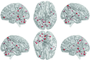
Gold Standard for Epilepsy/Tumor Surgery Coupled with Deep Learning Offers Independence to a Promising Functional Mapping Modality
Authors: Korostenskaja, M., RaviPrakash, H., Bagci, U., … , Elsayed, M., and Castillo, E.
Brain-Computer Interface Research, A State-of-the-Art Summary 7, Springer, 2019.
Publication Year: 2019
Artificial Intelligence as Another Set of Eyes in Breast Cancer Diagnosis
Authors: Anwar, SM, and Bagci, U.
Journal of Medical Artificial Intelligence - 2019
Publication Year: 2019


Prognostic Utility of F-18-NaF PET/CT Imaging for Fractures in Patients with Fibrous Dysplasia of Bone.
Authors: Papadakis, GZ., Manikis, GC, Karantanas, AH., Marias, K., Bagci, U., Florenzano, P., Collins, MT, Boyce, AM.
EJNMMI - 2019
Publication Year: 2019

MRI-guided, Transperineal Prostate Biopsy Using Fixed Coordinate Needle Guide: Initial Feasibility Study.
Authors: Kulkarni, P., Laha, S., Sikander, S., Cornell, H., Bagci, U., Burt, J., and Song, SS.
Design of Medical Devices Conference, 2019.
Publication Year: 2019
F-18-NaF Uptake by Fibrous Dysplasia Bone Lesions is Positively Associated with Bone Turnover Markers (BTMs)
Authors: Papadakis, GZ., Manikis, GC., Karantanas, AH., Marias, K., Bagci, U., Florenzano, P., Collins, MT, Boyce, AM.
EJNMMI - 2019
Publication Year: 2019
Colorectcal Polyp Diagnosis With Contemporary Artificial Intelligence
Authors: Kandel, P., Lalonde, R., Ciofoaia, V., Wallace, M., Bagci, U.
Gastrointestinal Endoscopy - 2019
Publication Year: 2019
Brown Adipose Tissue Activation Detected by Artificial Intelligence Assisted Radiomics: An Early Biomarker for Pancreatic Ductal Adenocarcinoma (PDAC)
Authors: Rodriguez, AC., Sharma, A., Tanner, I., Kandel, P., Lalonde, R., Livingston, D., Manoj, J., Raimondo, M., Wallace, M., Bagci, U.
Gastroenterology - 2019
Publication Year: 2019